6EZ8

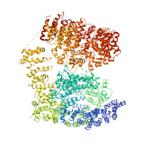
Human Huntingtin-HAP40 complex structure


Experimental Data Snapshot

  • Method: ELECTRON MICROSCOPY
  • Resolution: 4.00 Å
  • Aggregation State: PARTICLE 
  • Reconstruction Method: SINGLE PARTICLE 

wwPDB Validation   3D Report Full Report


This is version 2.1 of the entry. See complete history


Literature

The cryo-electron microscopy structure of huntingtin.

Guo, Q.Cheng, J.Seefelder, M.Engler, T.Pfeifer, G.Oeckl, P.Otto, M.Moser, F.Maurer, M.Pautsch, A.Baumeister, W.Fernandez-Busnadiego, R.Kochanek, S.

(2018) Nature 555: 117-120

  • DOI: https://doi.org/10.1038/nature25502
  • Primary Citation of Related Structures:  
    6EZ8

  • PubMed Abstract: 

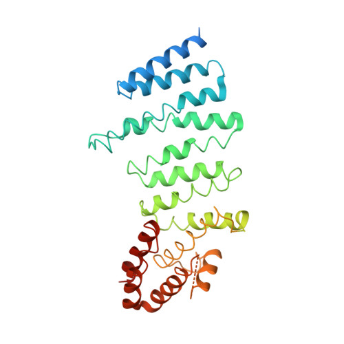
    Huntingtin (HTT) is a large (348 kDa) protein that is essential for embryonic development and is involved in diverse cellular activities such as vesicular transport, endocytosis, autophagy and the regulation of transcription. Although an integrative understanding of the biological functions of HTT is lacking, the large number of identified HTT interactors suggests that it serves as a protein-protein interaction hub. Furthermore, Huntington's disease is caused by a mutation in the HTT gene, resulting in a pathogenic expansion of a polyglutamine repeat at the amino terminus of HTT. However, only limited structural information regarding HTT is currently available. Here we use cryo-electron microscopy to determine the structure of full-length human HTT in a complex with HTT-associated protein 40 (HAP40; encoded by three F8A genes in humans) to an overall resolution of 4 Å. HTT is largely α-helical and consists of three major domains. The amino- and carboxy-terminal domains contain multiple HEAT (huntingtin, elongation factor 3, protein phosphatase 2A and lipid kinase TOR) repeats arranged in a solenoid fashion. These domains are connected by a smaller bridge domain containing different types of tandem repeats. HAP40 is also largely α-helical and has a tetratricopeptide repeat-like organization. HAP40 binds in a cleft and contacts the three HTT domains by hydrophobic and electrostatic interactions, thereby stabilizing the conformation of HTT. These data rationalize previous biochemical results and pave the way for improved understanding of the diverse cellular functions of HTT.


  • Organizational Affiliation

    Department of Molecular Structural Biology, Max Planck Institute of Biochemistry, 82152 Martinsried, Germany.


Macromolecules
Find similar proteins by:  (by identity cutoff)  |  3D Structure
Entity ID: 1
MoleculeChains Sequence LengthOrganismDetailsImage
Huntingtin3,138Homo sapiensMutation(s): 0 
Gene Names: HTTHDIT15
UniProt & NIH Common Fund Data Resources
Find proteins for P42858 (Homo sapiens)
Explore P42858 
Go to UniProtKB:  P42858
PHAROS:  P42858
GTEx:  ENSG00000197386 
Entity Groups  
Sequence Clusters30% Identity50% Identity70% Identity90% Identity95% Identity100% Identity
UniProt GroupP42858
Sequence Annotations
Expand
  • Reference Sequence
Find similar proteins by:  (by identity cutoff)  |  3D Structure
Entity ID: 2
MoleculeChains Sequence LengthOrganismDetailsImage
Factor VIII intron 22 protein371Homo sapiensMutation(s): 0 
Gene Names: F8A1F8AF8A2F8A3
UniProt & NIH Common Fund Data Resources
Find proteins for P23610 (Homo sapiens)
Explore P23610 
Go to UniProtKB:  P23610
GTEx:  ENSG00000277150 
Entity Groups  
Sequence Clusters30% Identity50% Identity70% Identity90% Identity95% Identity100% Identity
UniProt GroupP23610
Sequence Annotations
Expand
  • Reference Sequence
Experimental Data & Validation

Experimental Data

  • Method: ELECTRON MICROSCOPY
  • Resolution: 4.00 Å
  • Aggregation State: PARTICLE 
  • Reconstruction Method: SINGLE PARTICLE 
EM Software:
TaskSoftware PackageVersion
RECONSTRUCTIONRELION2.1

Structure Validation

View Full Validation Report


Entry History & Funding Information

Deposition Data


Funding OrganizationLocationGrant Number
European CommissionGermanyFP7 GA ERC-2012-SyG_318987-ToPAG
CHDIUnited StatesA12302
CHDI foundationGermany--

Revision History  (Full details and data files)

  • Version 1.0: 2018-02-21
    Type: Initial release
  • Version 1.1: 2018-03-07
    Changes: Database references, Other
  • Version 2.0: 2018-03-28
    Changes: Advisory, Atomic model, Data collection, Database references, Derived calculations, Polymer sequence, Source and taxonomy, Structure summary
  • Version 2.1: 2019-12-11
    Changes: Other