6EQT | pdb_00006eqt

CRYSTAL STRUCTURE OF THE HUMAN KINETOCHORE PROTEIN CENP-N


Experimental Data Snapshot

  • Method: X-RAY DIFFRACTION
  • Resolution: 2.73 Å
  • R-Value Free: 
    0.254 (Depositor), 0.256 (DCC) 
  • R-Value Work: 
    0.206 (Depositor), 0.207 (DCC) 
  • R-Value Observed: 
    0.208 (Depositor) 

wwPDB Validation 3D Report Full Report

Validation slider image for 6EQT

This is version 1.2 of the entry. See complete history

Literature

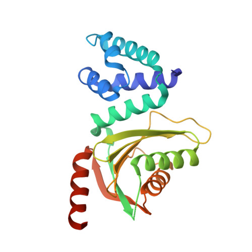
Decoding the centromeric nucleosome through CENP-N.

Pentakota, S.Zhou, K.Smith, C.Maffini, S.Petrovic, A.Morgan, G.P.Weir, J.R.Vetter, I.R.Musacchio, A.Luger, K.

(2017) Elife 6

  • DOI: https://doi.org/10.7554/eLife.33442
  • Primary Citation Related Structures: 
    6C0W, 6EQT

  • PubMed Abstract: 

    Centromere protein (CENP) A, a histone H3 variant, is a key epigenetic determinant of chromosome domains known as centromeres. Centromeres nucleate kinetochores, multi-subunit complexes that capture spindle microtubules to promote chromosome segregation during mitosis. Two kinetochore proteins, CENP-C and CENP-N, recognize CENP-A in the context of a rare CENP-A nucleosome. Here, we reveal the structural basis for the exquisite selectivity of CENP-N for centromeres. CENP-N uses charge and space complementarity to decode the L1 loop that is unique to CENP-A. It also engages in extensive interactions with a 15-base pair segment of the distorted nucleosomal DNA double helix, in a position predicted to exclude chromatin remodelling enzymes. Besides CENP-A, stable centromere recruitment of CENP-N requires a coincident interaction with a newly identified binding motif on nucleosome-bound CENP-C. Collectively, our studies clarify how CENP-N and CENP-C decode and stabilize the non-canonical CENP-A nucleosome to enforce epigenetic centromere specification and kinetochore assembly.


  • Organizational Affiliation
    • Department of Mechanistic Cell Biology, Max-Planck Institute of Molecular Physiology, Dortmund, Germany.

Macromolecule Content 

  • Total Structure Weight: 52.06 kDa 
  • Atom Count: 3,478 
  • Modeled Residue Count: 412 
  • Deposited Residue Count: 438 
  • Unique protein chains: 1

Macromolecules

Find similar proteins by:|  3D Structure
Entity ID: 1
MoleculeChains  Sequence LengthOrganismDetailsImage
Centromere protein N
A, B
219Homo sapiensMutation(s): 1 
Gene Names: CENPNC16orf60ICEN32BM-309
UniProt & NIH Common Fund Data Resources
Find proteins for Q96H22 (Homo sapiens)
Explore Q96H22 
Go to UniProtKB:  Q96H22
PHAROS:  Q96H22
GTEx:  ENSG00000166451 
Entity Groups
Sequence Clusters30% Identity50% Identity70% Identity90% Identity95% Identity100% Identity
UniProt GroupQ96H22
Sequence Annotations
Expand
Reference Sequence

Experimental Data & Validation

Experimental Data

  • Method: X-RAY DIFFRACTION
  • Resolution: 2.73 Å
  • R-Value Free:  0.254 (Depositor), 0.256 (DCC) 
  • R-Value Work:  0.206 (Depositor), 0.207 (DCC) 
  • R-Value Observed: 0.208 (Depositor) 
Space Group: P 41
Unit Cell:
Length ( Å )Angle ( ˚ )
a = 87.3α = 90
b = 87.3β = 90
c = 81.1γ = 90
Software Package:
Software NamePurpose
REFMACrefinement
XSCALEdata reduction
PDB_EXTRACTdata extraction
XSCALEdata scaling
PHENIXphasing

Structure Validation

View Full Validation Report


Entry History 

Deposition Data

Revision History  (Full details and data files)

  • Version 1.0: 2018-01-17
    Type: Initial release
  • Version 1.1: 2018-01-31
    Changes: Refinement description
  • Version 1.2: 2024-05-08
    Changes: Data collection, Database references