


Crystal structure of 7,8-diaminopelargonic acid synthase bound to inhibitor MAC13772
Brown, C.M., Zlitni, S., Chan, J., Brown, E.D., Junop, M.S.To be published.
Experimental Data Snapshot
Entity ID: 1 | |||||
|---|---|---|---|---|---|
| Molecule | Chains | Sequence Length | Organism | Details | Image |
| 7,8-diamino-pelargonic acid aminotransferase | 429 | Escherichia coli | Mutation(s): 0 Gene Names: bioA, EL75_3021, EL79_3111, EL80_3072 EC: 2.6.1.62 | 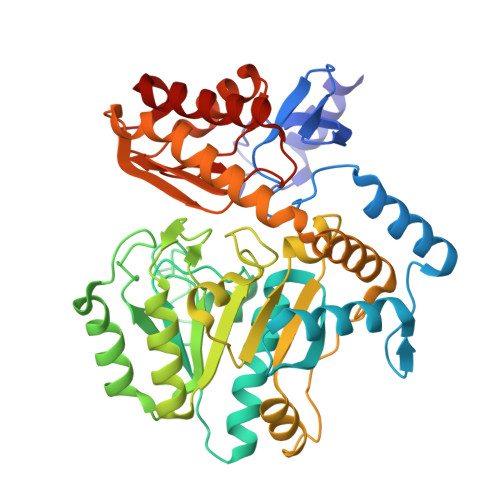 | |
UniProt | |||||
Entity Groups | |||||
| Sequence Clusters | 30% Identity50% Identity70% Identity90% Identity95% Identity100% Identity | ||||
| UniProt Group | P12995 | ||||
Sequence AnnotationsExpand | |||||
Reference Sequence | |||||
| Ligands 2 Unique | |||||
|---|---|---|---|---|---|
| ID | Chains | Name / Formula / InChI Key | 2D Diagram | 3D Interactions | |
| PLP Download:Ideal Coordinates CCD File | BA [auth G] DA [auth H] I [auth A] L [auth B] O [auth C] | PYRIDOXAL-5'-PHOSPHATE C8 H10 N O6 P NGVDGCNFYWLIFO-UHFFFAOYSA-N |  | ||
| J4J Download:Ideal Coordinates CCD File | AA [auth G] CA [auth G] EA [auth H] FA [auth H] J [auth A] | 2-[(2-nitrophenyl)sulfanyl]acetohydrazide C8 H9 N3 O3 S CZXFCCXLFQCYBK-UHFFFAOYSA-N |  | ||
| Length ( Å ) | Angle ( ˚ ) |
|---|---|
| a = 122.43 | α = 90 |
| b = 111.57 | β = 92.08 |
| c = 136.12 | γ = 90 |
| Software Name | Purpose |
|---|---|
| PHENIX | refinement |
| HKL-2000 | data reduction |
| HKL-2000 | data scaling |
| PHASER | phasing |
| Funding Organization | Location | Grant Number |
|---|---|---|
| Canadian Institutes of Health Research (CIHR) | Canada | -- |


