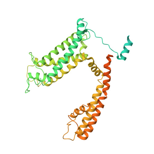
6EBM

The voltage-activated Kv1.2-2.1 paddle chimera channel in lipid nanodiscs, transmembrane domain of subunit alpha


Experimental Data Snapshot

  • Method: ELECTRON MICROSCOPY
  • Resolution: 4.00 Å
  • Aggregation State: PARTICLE 
  • Reconstruction Method: SINGLE PARTICLE 

Starting Model: experimental
View more details

wwPDB Validation   3D Report Full Report


This is version 1.1 of the entry. See complete history


Literature

Single-particle cryo-EM structure of a voltage-activated potassium channel in lipid nanodiscs.

Matthies, D.Bae, C.Toombes, G.E.Fox, T.Bartesaghi, A.Subramaniam, S.Swartz, K.J.

(2018) Elife 7

  • DOI: https://doi.org/10.7554/eLife.37558
  • Primary Citation of Related Structures:  
    6EBK, 6EBL, 6EBM

  • PubMed Abstract: 

    Voltage-activated potassium (Kv) channels open to conduct K + ions in response to membrane depolarization, and subsequently enter non-conducting states through distinct mechanisms of inactivation. X-ray structures of detergent-solubilized Kv channels appear to have captured an open state even though a non-conducting C-type inactivated state would predominate in membranes in the absence of a transmembrane voltage. However, structures for a voltage-activated ion channel in a lipid bilayer environment have not yet been reported. Here we report the structure of the Kv1.2-2.1 paddle chimera channel reconstituted into lipid nanodiscs using single-particle cryo-electron microscopy. At a resolution of ~3 Å for the cytosolic domain and ~4 Å for the transmembrane domain, the structure determined in nanodiscs is similar to the previously determined X-ray structure. Our findings show that large differences in structure between detergent and lipid bilayer environments are unlikely, and enable us to propose possible structural mechanisms for C-type inactivation.


  • Organizational Affiliation

    Laboratory of Cell Biology, Center for Cancer Research, National Cancer Institute, National Institutes of Health, Bethesda, United States.


Macromolecules
Find similar proteins by:  (by identity cutoff)  |  3D Structure
Entity ID: 1
MoleculeChains Sequence LengthOrganismDetailsImage
Potassium voltage-gated channel subfamily A member 2,Potassium voltage-gated channel subfamily B member 2 chimeraA [auth B],
B [auth D],
C [auth F],
D [auth H]
513Rattus norvegicusMutation(s): 0 
Gene Names: Kcna2Kcnb2
Membrane Entity: Yes 
UniProt
Find proteins for P63142 (Rattus norvegicus)
Explore P63142 
Go to UniProtKB:  P63142
Find proteins for Q63099 (Rattus norvegicus)
Explore Q63099 
Go to UniProtKB:  Q63099
Entity Groups  
Sequence Clusters30% Identity50% Identity70% Identity90% Identity95% Identity100% Identity
UniProt GroupsP63142Q63099
Sequence Annotations
Expand
  • Reference Sequence
Experimental Data & Validation

Experimental Data

  • Method: ELECTRON MICROSCOPY
  • Resolution: 4.00 Å
  • Aggregation State: PARTICLE 
  • Reconstruction Method: SINGLE PARTICLE 
EM Software:
TaskSoftware PackageVersion
RECONSTRUCTIONRELION2.1
MODEL REFINEMENTCoot
MODEL REFINEMENTPHENIX

Structure Validation

View Full Validation Report


Entry History 

Deposition Data

Revision History  (Full details and data files)

  • Version 1.0: 2018-08-22
    Type: Initial release
  • Version 1.1: 2024-03-13
    Changes: Data collection, Database references, Refinement description