6DQX | pdb_00006dqx

Actinobacillus ureae class Id ribonucleotide reductase alpha subunit


Experimental Data Snapshot

  • Method: X-RAY DIFFRACTION
  • Resolution: 1.76 Å
  • R-Value Free: 
    0.224 (Depositor), 0.233 (DCC) 
  • R-Value Work: 
    0.192 (Depositor), 0.202 (DCC) 
  • R-Value Observed: 
    0.193 (Depositor) 

Starting Model: experimental
View more details

wwPDB Validation 3D Report Full Report

Validation slider image for 6DQX

This is version 1.3 of the entry. See complete history

Literature

Structures of Class Id Ribonucleotide Reductase Catalytic Subunits Reveal a Minimal Architecture for Deoxynucleotide Biosynthesis.

Rose, H.R.Maggiolo, A.O.McBride, M.J.Palowitch, G.M.Pandelia, M.E.Davis, K.M.Yennawar, N.H.Boal, A.K.

(2019) Biochemistry 58: 1845-1860

  • DOI: https://doi.org/10.1021/acs.biochem.8b01252
  • Primary Citation Related Structures: 
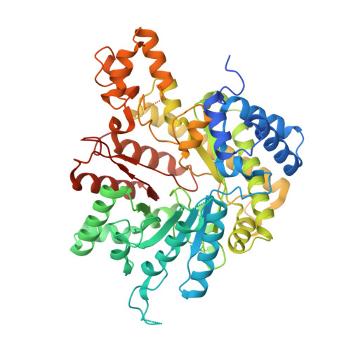
    6DQW, 6DQX

  • PubMed Abstract: 

    Class I ribonucleotide reductases (RNRs) share a common mechanism of nucleotide reduction in a catalytic α subunit. All RNRs initiate catalysis with a thiyl radical, generated in class I enzymes by a metallocofactor in a separate β subunit. Class Id RNRs use a simple mechanism of cofactor activation involving oxidation of a Mn II 2 cluster by free superoxide to yield a metal-based Mn III Mn IV oxidant. This simple cofactor assembly pathway suggests that class Id RNRs may be representative of the evolutionary precursors to more complex class Ia-c enzymes. X-ray crystal structures of two class Id α proteins from Flavobacterium johnsoniae ( Fj) and Actinobacillus ureae ( Au) reveal that this subunit is distinctly small. The enzyme completely lacks common N-terminal ATP-cone allosteric motifs that regulate overall activity, a process that normally occurs by dATP-induced formation of inhibitory quaternary structures to prevent productive β subunit association. Class Id RNR activity is insensitive to dATP in the Fj and Au enzymes evaluated here, as expected. However, the class Id α protein from Fj adopts higher-order structures, detected crystallographically and in solution. The Au enzyme does not exhibit these quaternary forms. Our study reveals structural similarity between bacterial class Id and eukaryotic class Ia α subunits in conservation of an internal auxiliary domain. Our findings with the Fj enzyme illustrate that nucleotide-independent higher-order quaternary structures can form in simple RNRs with truncated or missing allosteric motifs.


  • Organizational Affiliation
    • Department of Chemistry , The Pennsylvania State University , University Park , Pennsylvania 16802 , United States.

Macromolecule Content 

  • Total Structure Weight: 65.7 kDa 
  • Atom Count: 4,462 
  • Modeled Residue Count: 520 
  • Deposited Residue Count: 574 
  • Unique protein chains: 1

Macromolecules

Find similar proteins by:|  3D Structure
Entity ID: 1
MoleculeChains  Sequence LengthOrganismDetailsImage
Ribonucleoside-diphosphate reductase, alpha chain574Actinobacillus ureae ATCC 25976Mutation(s): 0 
Gene Names: HMPREF0027_1834
EC: 1.17.4.1
UniProt
Find proteins for E8KJ17 (Actinobacillus ureae ATCC 25976)
Explore E8KJ17 
Go to UniProtKB:  E8KJ17
Entity Groups
Sequence Clusters30% Identity50% Identity70% Identity90% Identity95% Identity100% Identity
UniProt GroupE8KJ17
Sequence Annotations
Expand
Reference Sequence

Small Molecules

Ligands 4 Unique
IDChains Name / Formula / InChI Key2D Diagram3D Interactions
GOL

Query on GOL


Download:Ideal Coordinates CCD File
J [auth A]GLYCEROL
C3 H8 O3
PEDCQBHIVMGVHV-UHFFFAOYSA-N
EDO

Query on EDO


Download:Ideal Coordinates CCD File
F [auth A],
G [auth A],
H [auth A],
I [auth A]
1,2-ETHANEDIOL
C2 H6 O2
LYCAIKOWRPUZTN-UHFFFAOYSA-N
CL

Query on CL


Download:Ideal Coordinates CCD File
B [auth A],
C [auth A]
CHLORIDE ION
Cl
VEXZGXHMUGYJMC-UHFFFAOYSA-M
MG

Query on MG


Download:Ideal Coordinates CCD File
D [auth A],
E [auth A]
MAGNESIUM ION
Mg
JLVVSXFLKOJNIY-UHFFFAOYSA-N

Experimental Data & Validation

Experimental Data

  • Method: X-RAY DIFFRACTION
  • Resolution: 1.76 Å
  • R-Value Free:  0.224 (Depositor), 0.233 (DCC) 
  • R-Value Work:  0.192 (Depositor), 0.202 (DCC) 
  • R-Value Observed: 0.193 (Depositor) 
Space Group: P 42 21 2
Unit Cell:
Length ( Å )Angle ( ˚ )
a = 97.984α = 90
b = 97.984β = 90
c = 132.005γ = 90
Software Package:
Software NamePurpose
REFMACrefinement
HKL-2000data scaling
PDB_EXTRACTdata extraction
HKL-2000data reduction
PHASERphasing

Structure Validation

View Full Validation Report


Entry History 

& Funding Information

Deposition Data


Funding OrganizationLocationGrant Number
National Institutes of Health/National Institute of General Medical Sciences (NIH/NIGMS)United StatesGM119707

Revision History  (Full details and data files)

  • Version 1.0: 2019-04-17
    Type: Initial release
  • Version 1.1: 2019-04-24
    Changes: Data collection, Database references
  • Version 1.2: 2020-01-01
    Changes: Author supporting evidence
  • Version 1.3: 2023-10-11
    Changes: Data collection, Database references, Derived calculations, Refinement description