


Crystal structure of DNA polymerase III subunit beta from Mycobacterium marinum in complex with a natural product
Bowatte, K., Conrady, D.G., Abendroth, J., Lorimer, D.D., Edwards, T.E.To be published.
Experimental Data Snapshot
Starting Model: experimental
View more details
wwPDB Validation 3D Report Full Report

Entity ID: 1 | |||||
|---|---|---|---|---|---|
| Molecule | Chains | Sequence Length | Organism | Details | Image |
| Beta sliding clamp | 410 | Mycobacterium marinum M | Mutation(s): 0 Gene Names: dnaN, MMAR_0002 | 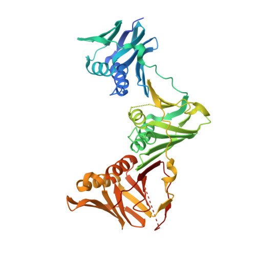 | |
UniProt | |||||
Entity Groups | |||||
| Sequence Clusters | 30% Identity50% Identity70% Identity90% Identity95% Identity100% Identity | ||||
| UniProt Group | B2HI47 | ||||
Sequence AnnotationsExpand | |||||
Reference Sequence | |||||
Entity ID: 2 | |||||
|---|---|---|---|---|---|
| Molecule | Chains | Sequence Length | Organism | Details | Image |
| Natural product peptide | 11 | Streptomyces muensis | Mutation(s): 0 | 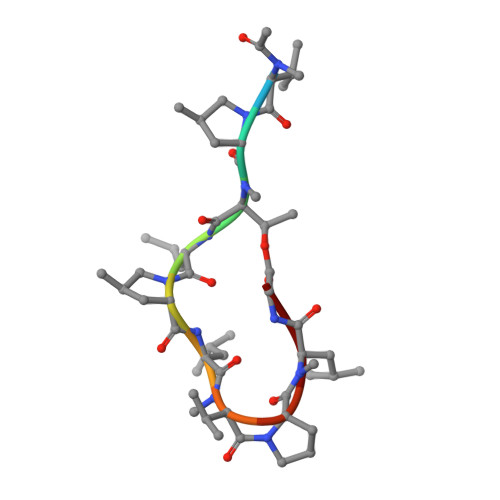 | |
| Ligands 1 Unique | |||||
|---|---|---|---|---|---|
| ID | Chains | Name / Formula / InChI Key | 2D Diagram | 3D Interactions | |
| EDO Download:Ideal Coordinates CCD File | E [auth B] | 1,2-ETHANEDIOL C2 H6 O2 LYCAIKOWRPUZTN-UHFFFAOYSA-N |  | ||
| Modified Residues 3 Unique | |||||
|---|---|---|---|---|---|
| ID | Chains | Type | Formula | 2D Diagram | Parent |
| MLU Query on MLU | C, D | D-PEPTIDE LINKING | C7 H15 N O2 |  | -- |
| MP8 Query on MP8 | C, D | L-PEPTIDE LINKING | C6 H11 N O2 |  | PRO |
| MVA Query on MVA | C, D | L-PEPTIDE LINKING | C6 H13 N O2 |  | VAL |
| NZC Query on NZC | C, D | L-PEPTIDE LINKING | C5 H11 N O3 |  | THR |
| Entity ID: 2 | |||||
|---|---|---|---|---|---|
| ID | Chains | Name | Type/Class | 2D Diagram | 3D Interactions |
| PRD_002311 Query on PRD_002311 | C, D | ACE-MVA-MP8-NZC-LEU-MP8-LEU-MVA-PRO-MLU-GLY | Peptide-like / Inhibitor |  | |
| Length ( Å ) | Angle ( ˚ ) |
|---|---|
| a = 80.09 | α = 90 |
| b = 76.76 | β = 117.53 |
| c = 80.79 | γ = 90 |
| Software Name | Purpose |
|---|---|
| PHENIX | refinement |
| XSCALE | data scaling |
| PDB_EXTRACT | data extraction |
| XDS | data reduction |
| PHASER | phasing |


