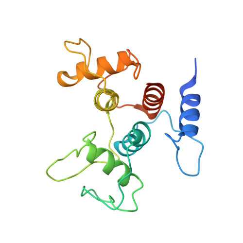
6CWN

Crystal structure of SpaA-SLH/G109A in complex with 4,6-Pyr-beta-D-ManNAcOMe


Experimental Data Snapshot

  • Method: X-RAY DIFFRACTION
  • Resolution: 1.53 Å
  • R-Value Free: 0.151 
  • R-Value Work: 0.113 
  • R-Value Observed: 0.115 

wwPDB Validation   3D Report Full Report


Ligand Structure Quality Assessment 


This is version 1.3 of the entry. See complete history


Literature

Structural basis of cell wall anchoring by SLH domains in Paenibacillus alvei.

Blackler, R.J.Lopez-Guzman, A.Hager, F.F.Janesch, B.Martinz, G.Gagnon, S.M.L.Haji-Ghassemi, O.Kosma, P.Messner, P.Schaffer, C.Evans, S.V.

(2018) Nat Commun 9: 3120-3120

  • DOI: https://doi.org/10.1038/s41467-018-05471-3
  • Primary Citation of Related Structures:  
    6CWC, 6CWF, 6CWH, 6CWI, 6CWL, 6CWM, 6CWN, 6CWR

  • PubMed Abstract: 

    Self-assembling protein surface (S-) layers are common cell envelope structures of prokaryotes and have critical roles from structural maintenance to virulence. S-layers of Gram-positive bacteria are often attached through the interaction of S-layer homology (SLH) domain trimers with peptidoglycan-linked secondary cell wall polymers (SCWPs). Here we present an in-depth characterization of this interaction, with co-crystal structures of the three consecutive SLH domains from the Paenibacillus alvei S-layer protein SpaA with defined SCWP ligands. The most highly conserved SLH domain residue SLH-Gly29 is shown to enable a peptide backbone flip essential for SCWP binding in both biophysical and cellular experiments. Furthermore, we find that a significant domain movement mediates binding by two different sites in the SLH domain trimer, which may allow anchoring readjustment to relieve S-layer strain caused by cell growth and division.


  • Organizational Affiliation

    Department of Biochemistry and Microbiology, University of Victoria, Victoria, BC, V8W 3P6, Canada.


Macromolecules
Find similar proteins by:  (by identity cutoff)  |  3D Structure
Entity ID: 1
MoleculeChains Sequence LengthOrganismDetailsImage
Surface (S-) layer glycoprotein182Paenibacillus alveiMutation(s): 1 
Gene Names: spaA
UniProt
Find proteins for C1JZ07 (Paenibacillus alvei)
Explore C1JZ07 
Go to UniProtKB:  C1JZ07
Entity Groups  
Sequence Clusters30% Identity50% Identity70% Identity90% Identity95% Identity100% Identity
UniProt GroupC1JZ07
Sequence Annotations
Expand
  • Reference Sequence
Experimental Data & Validation

Experimental Data

  • Method: X-RAY DIFFRACTION
  • Resolution: 1.53 Å
  • R-Value Free: 0.151 
  • R-Value Work: 0.113 
  • R-Value Observed: 0.115 
  • Space Group: C 1 2 1
Unit Cell:
Length ( Å )Angle ( ˚ )
a = 93.383α = 90
b = 36.254β = 103.74
c = 56.896γ = 90
Software Package:
Software NamePurpose
HKL-2000data scaling
REFMACrefinement
PDB_EXTRACTdata extraction
HKL-2000data reduction
PHASERphasing

Structure Validation

View Full Validation Report


Ligand Structure Quality Assessment 


Entry History & Funding Information

Deposition Data


Funding OrganizationLocationGrant Number
Natural Sciences and Engineering Research Council (NSERC, Canada)CanadaCGSD3-426678-2012
Austrian Science FundAustriaP22791-B12
Austrian Science FundAustriaP27374-B22

Revision History  (Full details and data files)

  • Version 1.0: 2018-08-15
    Type: Initial release
  • Version 1.1: 2018-08-22
    Changes: Data collection, Database references
  • Version 1.2: 2020-01-08
    Changes: Author supporting evidence
  • Version 1.3: 2024-03-13
    Changes: Data collection, Database references, Refinement description