6C5W

Crystal structure of the mitochondrial calcium uniporter


Experimental Data Snapshot

  • Method: X-RAY DIFFRACTION
  • Resolution: 3.10 Å
  • R-Value Free: 0.299 
  • R-Value Work: 0.256 
  • R-Value Observed: 0.258 

wwPDB Validation   3D Report Full Report


This is version 1.4 of the entry. See complete history


Literature

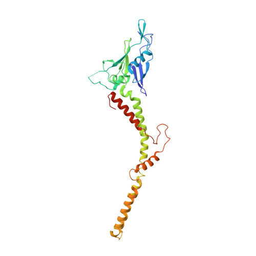
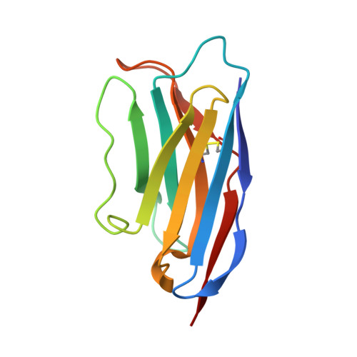
X-ray and cryo-EM structures of the mitochondrial calcium uniporter.

Fan, C.Fan, M.Orlando, B.J.Fastman, N.M.Zhang, J.Xu, Y.Chambers, M.G.Xu, X.Perry, K.Liao, M.Feng, L.

(2018) Nature 559: 575-579

  • DOI: https://doi.org/10.1038/s41586-018-0330-9
  • Primary Citation of Related Structures:  
    6C5R, 6C5W

  • PubMed Abstract: 

    Mitochondrial calcium uptake is critical for regulating ATP production, intracellular calcium signalling, and cell death. This uptake is mediated by a highly selective calcium channel called the mitochondrial calcium uniporter (MCU). Here, we determined the structures of the pore-forming MCU proteins from two fungi by X-ray crystallography and single-particle cryo-electron microscopy. The stoichiometry, overall architecture, and individual subunit structure differed markedly from those described in the recent nuclear magnetic resonance structure of Caenorhabditis elegans MCU. We observed a dimer-of-dimer architecture across species and chemical environments, which was corroborated by biochemical experiments. Structural analyses and functional characterization uncovered the roles of key residues in the pore. These results reveal a new ion channel architecture, provide insights into calcium coordination, selectivity and conduction, and establish a structural framework for understanding the mechanism of mitochondrial calcium uniporter function.


  • Organizational Affiliation

    Department of Molecular and Cellular Physiology, Stanford University School of Medicine, Stanford, CA, USA.


Macromolecules
Find similar proteins by:  (by identity cutoff)  |  3D Structure
Entity ID: 1
MoleculeChains Sequence LengthOrganismDetailsImage
calcium uniporter
A, B
312Metarhizium acridum CQMa 102Mutation(s): 0 
Gene Names: MAC_01752
Membrane Entity: Yes 
UniProt
Find proteins for E9DVV4 (Metarhizium acridum (strain CQMa 102))
Explore E9DVV4 
Go to UniProtKB:  E9DVV4
Entity Groups  
Sequence Clusters30% Identity50% Identity70% Identity90% Identity95% Identity100% Identity
UniProt GroupE9DVV4
Sequence Annotations
Expand
  • Reference Sequence
Find similar proteins by:  (by identity cutoff)  |  3D Structure
Entity ID: 2
MoleculeChains Sequence LengthOrganismDetailsImage
nanobodyC,
D [auth E]
117unidentifiedMutation(s): 0 
Entity Groups  
Sequence Clusters30% Identity50% Identity70% Identity90% Identity95% Identity100% Identity
Sequence Annotations
Expand
  • Reference Sequence
Small Molecules
Ligands 1 Unique
IDChains Name / Formula / InChI Key2D Diagram3D Interactions
CA
Query on CA

Download Ideal Coordinates CCD File 
E [auth A]CALCIUM ION
Ca
BHPQYMZQTOCNFJ-UHFFFAOYSA-N
Experimental Data & Validation

Experimental Data

  • Method: X-RAY DIFFRACTION
  • Resolution: 3.10 Å
  • R-Value Free: 0.299 
  • R-Value Work: 0.256 
  • R-Value Observed: 0.258 
  • Space Group: C 2 2 21
Unit Cell:
Length ( Å )Angle ( ˚ )
a = 88.02α = 90
b = 290.45β = 90
c = 126.12γ = 90
Software Package:
Software NamePurpose
phenix.refinerefinement
PHENIXrefinement
HKL-2000data reduction
HKL-2000data scaling
PHENIXphasing

Structure Validation

View Full Validation Report


Entry History 

Deposition Data

Revision History  (Full details and data files)

  • Version 1.0: 2018-07-11
    Type: Initial release
  • Version 1.1: 2018-07-25
    Changes: Data collection, Database references
  • Version 1.2: 2018-08-08
    Changes: Data collection, Database references
  • Version 1.3: 2018-10-03
    Changes: Data collection, Source and taxonomy
  • Version 1.4: 2019-04-24
    Changes: Author supporting evidence, Data collection