6C5W | pdb_00006c5w

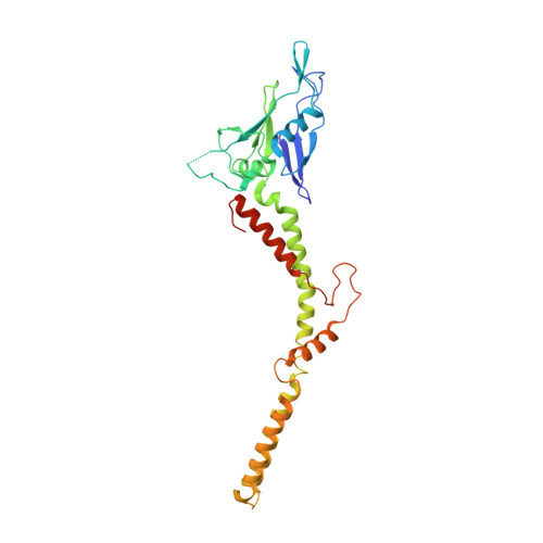
Crystal structure of the mitochondrial calcium uniporter


Experimental Data Snapshot

  • Method: X-RAY DIFFRACTION
  • Resolution: 3.10 Å
  • R-Value Free: 
    0.299 (Depositor), 0.302 (DCC) 
  • R-Value Work: 
    0.256 (Depositor), 0.257 (DCC) 
  • R-Value Observed: 
    0.258 (Depositor) 

wwPDB Validation 3D Report Full Report

Validation slider image for 6C5W

This is version 1.5 of the entry. See complete history. 

Literature

X-ray and cryo-EM structures of the mitochondrial calcium uniporter.

Fan, C., Fan, M., Orlando, B.J., Fastman, N.M., Zhang, J., Xu, Y., Chambers, M.G., Xu, X., Perry, K., Liao, M., Feng, L.

(2018) Nature 559: 575-579

  • DOI: https://doi.org/10.1038/s41586-018-0330-9
  • Primary Citation Related Structures: 
    6C5R, 6C5W

  • PubMed Abstract: 

    Mitochondrial calcium uptake is critical for regulating ATP production, intracellular calcium signalling, and cell death. This uptake is mediated by a highly selective calcium channel called the mitochondrial calcium uniporter (MCU). Here, we determined the structures of the pore-forming MCU proteins from two fungi by X-ray crystallography and single-particle cryo-electron microscopy. The stoichiometry, overall architecture, and individual subunit structure differed markedly from those described in the recent nuclear magnetic resonance structure of Caenorhabditis elegans MCU. We observed a dimer-of-dimer architecture across species and chemical environments, which was corroborated by biochemical experiments. Structural analyses and functional characterization uncovered the roles of key residues in the pore. These results reveal a new ion channel architecture, provide insights into calcium coordination, selectivity and conduction, and establish a structural framework for understanding the mechanism of mitochondrial calcium uniporter function.


  • Organizational Affiliation: 
    • Department of Molecular and Cellular Physiology, Stanford University School of Medicine, Stanford, CA, USA.

Macromolecule Content 

  • Total Structure Weight: 96.53 kDa 
  • Atom Count: 5,745 
  • Modeled Residue Count: 736 
  • Deposited Residue Count: 858 
  • Unique protein chains: 2

Macromolecules

Find similar proteins by:|  3D Structure
Entity ID: 1
MoleculeChains  Sequence LengthOrganismDetailsImage
calcium uniporter
A, B
312Metarhizium acridum CQMa 102Mutation(s): 0 
Gene Names: MAC_01752
Membrane Entity: Yes 
UniProt
Find proteins for E9DVV4 (Metarhizium acridum (strain CQMa 102))
Explore E9DVV4 
Go to UniProtKB:  E9DVV4
Entity Groups
Sequence Clusters30% Identity50% Identity70% Identity90% Identity95% Identity100% Identity
UniProt GroupE9DVV4
Sequence Annotations
Expand
Reference Sequence
Find similar proteins by:|  3D Structure
Entity ID: 2
MoleculeChains  Sequence LengthOrganismDetailsImage
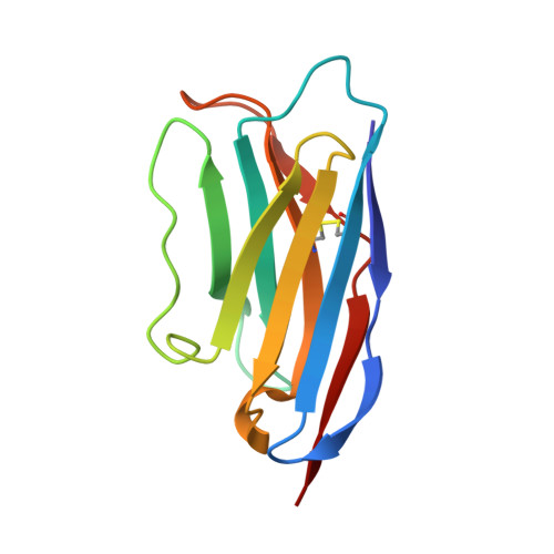
nanobodyC,
D [auth E]
117unidentifiedMutation(s): 0 
Entity Groups
Sequence Clusters30% Identity50% Identity70% Identity90% Identity95% Identity100% Identity
Sequence Annotations
Expand
Reference Sequence

Small Molecules

Ligands 1 Unique
IDChains Name / Formula / InChI Key2D Diagram3D Interactions
CA

Query on CA


Download:Ideal Coordinates CCD File
E [auth A]CALCIUM ION
Ca
BHPQYMZQTOCNFJ-UHFFFAOYSA-N

Experimental Data & Validation

Experimental Data

  • Method: X-RAY DIFFRACTION
  • Resolution: 3.10 Å
  • R-Value Free:  0.299 (Depositor), 0.302 (DCC) 
  • R-Value Work:  0.256 (Depositor), 0.257 (DCC) 
  • R-Value Observed: 0.258 (Depositor) 
Space Group: C 2 2 21
Unit Cell:
Length ( Å )Angle ( ˚ )
a = 88.02α = 90
b = 290.45β = 90
c = 126.12γ = 90
Software Package:
Software NamePurpose
phenix.refinerefinement
PHENIXrefinement
HKL-2000data reduction
HKL-2000data scaling
PHENIXphasing

Structure Validation

View Full Validation Report


Entry History 

Deposition Data

Revision History  (Full details and data files)

  • Version 1.0: 2018-07-11
    Type: Initial release
  • Version 1.1: 2018-07-25
    Changes: Data collection, Database references
  • Version 1.2: 2018-08-08
    Changes: Data collection, Database references
  • Version 1.3: 2018-10-03
    Changes: Data collection, Source and taxonomy
  • Version 1.4: 2019-04-24
    Changes: Author supporting evidence, Data collection
  • Version 1.5: 2024-11-20
    Changes: Data collection, Database references, Derived calculations, Structure summary