6BYL

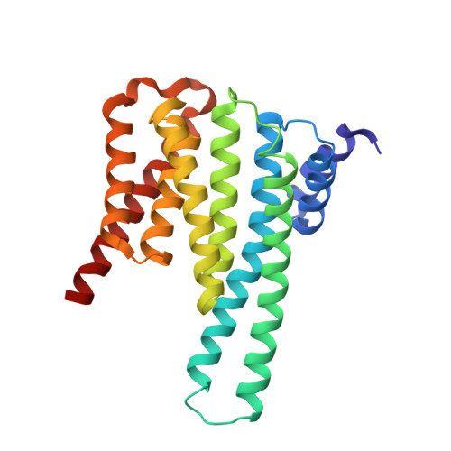
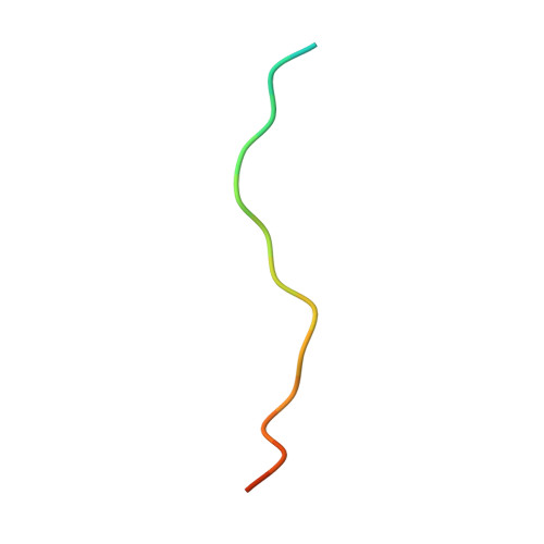
Structure of 14-3-3 gamma bound to O-GlcNAcylated thr peptide


Experimental Data Snapshot

  • Method: X-RAY DIFFRACTION
  • Resolution: 3.35 Å
  • R-Value Free: 
    0.285 (Depositor), 0.290 (DCC) 
  • R-Value Work: 
    0.234 (Depositor), 0.240 (DCC) 
  • R-Value Observed: 
    0.237 (Depositor) 

Starting Model: experimental
View more details

wwPDB Validation   3D Report Full Report


Ligand Structure Quality Assessment 

Created with Raphaël 2.3.0Worse 01 BetterLigand structure goodness of fit to experimental dataBest fitted NAGClick on this verticalbar to view details

This is version 1.4 of the entry. See complete history


Literature

Structural basis of O-GlcNAc recognition by mammalian 14-3-3 proteins.

Toleman, C.A.Schumacher, M.A.Yu, S.H.Zeng, W.Cox, N.J.Smith, T.J.Soderblom, E.J.Wands, A.M.Kohler, J.J.Boyce, M.

(2018) Proc Natl Acad Sci U S A 115: 5956-5961

  • DOI: https://doi.org/10.1073/pnas.1722437115
  • Primary Citation of Related Structures:  
    6BYJ, 6BYK, 6BYL, 6BZD

  • PubMed Abstract: 

    O-GlcNAc is an intracellular posttranslational modification that governs myriad cell biological processes and is dysregulated in human diseases. Despite this broad pathophysiological significance, the biochemical effects of most O-GlcNAcylation events remain uncharacterized. One prevalent hypothesis is that O-GlcNAc moieties may be recognized by "reader" proteins to effect downstream signaling. However, no general O-GlcNAc readers have been identified, leaving a considerable gap in the field. To elucidate O-GlcNAc signaling mechanisms, we devised a biochemical screen for candidate O-GlcNAc reader proteins. We identified several human proteins, including 14-3-3 isoforms, that bind O-GlcNAc directly and selectively. We demonstrate that 14-3-3 proteins bind O-GlcNAc moieties in human cells, and we present the structures of 14-3-3β/α and γ bound to glycopeptides, providing biophysical insights into O-GlcNAc-mediated protein-protein interactions. Because 14-3-3 proteins also bind to phospho-serine and phospho-threonine, they may integrate information from O-GlcNAc and O-phosphate signaling pathways to regulate numerous physiological functions.


  • Organizational Affiliation

    Department of Biochemistry, Duke University School of Medicine, Durham, NC 27710.


Macromolecules
Find similar proteins by:  (by identity cutoff)  |  3D Structure
Entity ID: 1
MoleculeChains Sequence LengthOrganismDetailsImage
14-3-3 protein gamma
A, B, C, D, E
240Homo sapiensMutation(s): 0 
Gene Names: YWHAG
UniProt & NIH Common Fund Data Resources
Find proteins for P61981 (Homo sapiens)
Explore P61981 
Go to UniProtKB:  P61981
PHAROS:  P61981
GTEx:  ENSG00000170027 
Entity Groups  
Sequence Clusters30% Identity50% Identity70% Identity90% Identity95% Identity100% Identity
UniProt GroupP61981
Sequence Annotations
Expand
  • Reference Sequence

Find similar proteins by:  Sequence   |   3D Structure  

Entity ID: 2
MoleculeChains Sequence LengthOrganismDetailsImage
TSASTTVPVTTATTTTTSTW O-GlcNac peptideG,
H [auth P],
I [auth T]
20synthetic constructMutation(s): 0 
Entity Groups  
Sequence Clusters30% Identity50% Identity70% Identity90% Identity95% Identity100% Identity
Glycosylation
Glycosylation Sites: 1
Sequence Annotations
Expand
  • Reference Sequence
Experimental Data & Validation

Experimental Data

  • Method: X-RAY DIFFRACTION
  • Resolution: 3.35 Å
  • R-Value Free:  0.285 (Depositor), 0.290 (DCC) 
  • R-Value Work:  0.234 (Depositor), 0.240 (DCC) 
  • R-Value Observed: 0.237 (Depositor) 
Space Group: P 41 21 2
Unit Cell:
Length ( Å )Angle ( ˚ )
a = 121.2α = 90
b = 121.2β = 90
c = 310.9γ = 90
Software Package:
Software NamePurpose
PHENIXrefinement
MOSFLMdata collection
SCALAdata scaling
MOLREPphasing
PDB_EXTRACTdata extraction
MOSFLMdata reduction

Structure Validation

View Full Validation Report


Ligand Structure Quality Assessment 

Created with Raphaël 2.3.0Worse 01 BetterLigand structure goodness of fit to experimental dataBest fitted NAGClick on this verticalbar to view details

Entry History 

Deposition Data

Revision History  (Full details and data files)

  • Version 1.0: 2018-05-09
    Type: Initial release
  • Version 1.1: 2019-05-22
    Changes: Data collection, Database references
  • Version 1.2: 2020-07-29
    Type: Remediation
    Reason: Carbohydrate remediation
    Changes: Data collection, Derived calculations, Structure summary
  • Version 1.3: 2023-10-04
    Changes: Data collection, Database references, Refinement description, Structure summary
  • Version 1.4: 2024-11-13
    Changes: Structure summary