6AU2 | pdb_00006au2

Crystal structure of SETDB1 Tudor domain with aryl triazole fragments


Experimental Data Snapshot

  • Method: X-RAY DIFFRACTION
  • Resolution: 1.63 Å
  • R-Value Free: 
    0.208 (Depositor), 0.216 (DCC) 
  • R-Value Work: 
    0.189 (Depositor), 0.201 (DCC) 
  • R-Value Observed: 
    0.189 (Depositor) 

wwPDB Validation 3D Report Full Report

Validation slider image for 6AU2

Ligand Structure Quality Assessment 


This is version 1.2 of the entry. See complete history

Literature

Identification and characterization of the first fragment hits for SETDB1 Tudor domain.

Mader, P.Mendoza-Sanchez, R.Iqbal, A.Dong, A.Dobrovetsky, E.Corless, V.B.Liew, S.K.Houliston, S.R.De Freitas, R.F.Smil, D.Sena, C.C.D.Kennedy, S.Diaz, D.B.Wu, H.Dombrovski, L.Allali-Hassani, A.Min, J.Schapira, M.Vedadi, M.Brown, P.J.Santhakumar, V.Yudin, A.K.Arrowsmith, C.H.

(2019) Bioorg Med Chem 27: 3866-3878

  • DOI: https://doi.org/10.1016/j.bmc.2019.07.020
  • Primary Citation Related Structures: 
    6AU2

  • PubMed Abstract: 

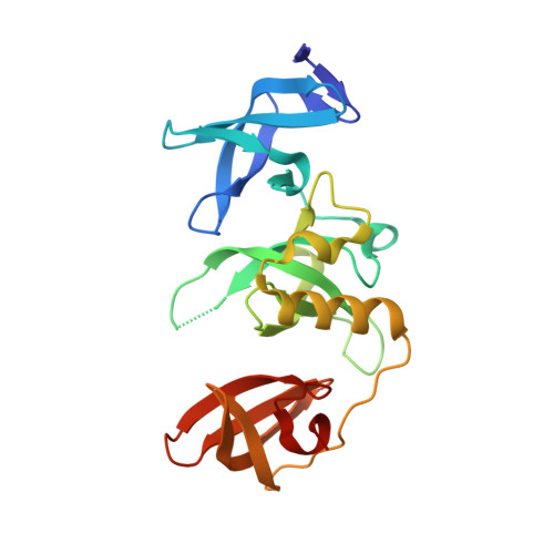
    SET domain bifurcated protein 1 (SETDB1) is a human histone-lysine methyltransferase which is amplified in human cancers and was shown to be crucial in the growth of non-small and small cell lung carcinoma. In addition to its catalytic domain, SETDB1 harbors a unique tandem tudor domain which recognizes histone sequences containing both methylated and acetylated lysines, and likely contributes to its localization on chromatin. Using X-ray crystallography and NMR spectroscopy fragment screening approaches, we have identified the first small molecule fragment hits that bind to histone peptide binding groove of the Tandem Tudor Domain (TTD) of SETDB1. Herein, we describe the binding modes of these fragments and analogues and the biophysical characterization of key compounds. These confirmed small molecule fragments will inform the development of potent antagonists of SETDB1 interaction with histones.


  • Organizational Affiliation
    • Structural Genomics Consortium, University of Toronto, Toronto, Canada.

Macromolecule Content 

  • Total Structure Weight: 26.1 kDa 
  • Atom Count: 2,058 
  • Modeled Residue Count: 210 
  • Deposited Residue Count: 213 
  • Unique protein chains: 1

Macromolecules

Find similar proteins by:|  3D Structure
Entity ID: 1
MoleculeChains  Sequence LengthOrganismDetailsImage
Histone-lysine N-methyltransferase SETDB1213Homo sapiensMutation(s): 0 
Gene Names: SETDB1KIAA0067KMT1E
EC: 2.1.1.43 (PDB Primary Data), 2.1.1.366 (UniProt)
UniProt & NIH Common Fund Data Resources
Find proteins for Q15047 (Homo sapiens)
Explore Q15047 
Go to UniProtKB:  Q15047
PHAROS:  Q15047
GTEx:  ENSG00000143379 
Entity Groups
Sequence Clusters30% Identity50% Identity70% Identity90% Identity95% Identity100% Identity
UniProt GroupQ15047
Sequence Annotations
Expand
Reference Sequence

Small Molecules

Ligands 5 Unique
IDChains Name / Formula / InChI Key2D Diagram3D Interactions
CGJ

Query on CGJ


Download:Ideal Coordinates CCD File
B [auth A]1-methyl-4H,6H-[1,2,4]triazolo[4,3-a][4,1]benzoxazepine
C11 H11 N3 O
HEEIZXCFEVISIX-UHFFFAOYSA-N
SO4

Query on SO4


Download:Ideal Coordinates CCD File
E [auth A],
F [auth A],
G [auth A],
H [auth A],
I [auth A]
SULFATE ION
O4 S
QAOWNCQODCNURD-UHFFFAOYSA-L
BME

Query on BME


Download:Ideal Coordinates CCD File
C [auth A],
D [auth A]
BETA-MERCAPTOETHANOL
C2 H6 O S
DGVVWUTYPXICAM-UHFFFAOYSA-N
EDO

Query on EDO


Download:Ideal Coordinates CCD File
J [auth A]
K [auth A]
L [auth A]
M [auth A]
N [auth A]
J [auth A],
K [auth A],
L [auth A],
M [auth A],
N [auth A],
O [auth A]
1,2-ETHANEDIOL
C2 H6 O2
LYCAIKOWRPUZTN-UHFFFAOYSA-N
UNX

Query on UNX


Download:Ideal Coordinates CCD File
AA [auth A]
BA [auth A]
P [auth A]
Q [auth A]
R [auth A]
AA [auth A],
BA [auth A],
P [auth A],
Q [auth A],
R [auth A],
S [auth A],
T [auth A],
U [auth A],
V [auth A],
W [auth A],
X [auth A],
Y [auth A],
Z [auth A]
UNKNOWN ATOM OR ION
X

Experimental Data & Validation

Experimental Data

  • Method: X-RAY DIFFRACTION
  • Resolution: 1.63 Å
  • R-Value Free:  0.208 (Depositor), 0.216 (DCC) 
  • R-Value Work:  0.189 (Depositor), 0.201 (DCC) 
  • R-Value Observed: 0.189 (Depositor) 
Space Group: P 21 21 21
Unit Cell:
Length ( Å )Angle ( ˚ )
a = 55.202α = 90
b = 63.273β = 90
c = 69.859γ = 90
Software Package:
Software NamePurpose
SCALEPACKdata scaling
REFMACrefinement
PDB_EXTRACTdata extraction
Cootmodel building
HKL-3000data reduction

Structure Validation

View Full Validation Report


Ligand Structure Quality Assessment 


Entry History 

Revision History  (Full details and data files)

  • Version 1.0: 2017-10-11
    Type: Initial release
  • Version 1.1: 2019-10-16
    Changes: Data collection, Database references
  • Version 1.2: 2025-04-02
    Changes: Data collection, Database references, Structure summary