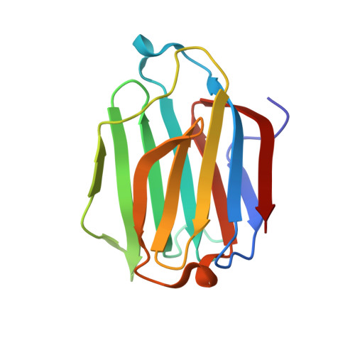
6A62

Placental protein 13/galectin-13 variant R53HH57RD33G with Lactose


Experimental Data Snapshot

  • Method: X-RAY DIFFRACTION
  • Resolution: 2.03 Å
  • R-Value Free: 0.215 
  • R-Value Work: 0.164 
  • R-Value Observed: 0.169 

wwPDB Validation   3D Report Full Report


This is version 2.1 of the entry. See complete history


Literature

Resetting the ligand binding site of placental protein 13/galectin-13 recovers its ability to bind lactose

Su, J.Cui, L.Si, Y.Song, C.Li, Y.Yang, T.Wang, H.Mayo, K.H.Tai, G.Zhou, Y.

(2018) Biosci Rep 38

  • DOI: https://doi.org/10.1042/BSR20181787
  • Primary Citation of Related Structures:  
    6A62, 6A63, 6A64, 6A65, 6A66

  • PubMed Abstract: 

    Placental protein 13/galectin-13 (Gal-13) is highly expressed in placenta, where its lower expression is related to pre-eclampsia. Recently, the crystal structures of wild-type Gal-13 and its variant R53H at high resolution were solved. The crystallographic and biochemical results showed that Gal-13 and R53H could not bind lactose. Here, we used site-directed mutagenesis to re-engineer the ligand binding site of wild-type Gal-13, so that it could bind lactose. Of six newly engineered mutants, we were able to solve the crystal structures of four of them. Three variants (R53HH57R, R53HH57RD33G and R53HR55NH57RD33G had the same two mutations (R53 to H, and H57 to R) and were able to bind lactose in the crystal, indicating that these mutations were sufficient for recovering the ability of Gal-13 to bind lactose. Moreover, the structures of R53H and R53HR55N show that these variants could co-crystallize with a molecule of Tris. Surprisingly, although these variants, as well as wild-type Gal-13, could all induce hemagglutination, high concentrations of lactose could not inhibit agglutination, nor could they bind to lactose-modified Sepharose 6b beads. Overall, our results indicate that Gal-3 is not a normal galectin, which could not bind to β-galactosides. Lastly, the distribution of EGFP-tagged wild-type Gal-13 and its variants in HeLa cells showed that they are concentrated in the nucleus and could be co-localized within filamentary materials, possibly actin.


  • Organizational Affiliation

    Jilin Province Key Laboratory for Chemistry and Biology of Natural Drugs in Changbai Mountain, School of Life Sciences, Northeast Normal University, Changchun 130024, China sujy100@nenu.edu.cn zhouyf383@nenu.edu.cn.


Macromolecules
Find similar proteins by:  (by identity cutoff)  |  3D Structure
Entity ID: 1
MoleculeChains Sequence LengthOrganismDetailsImage
Galactoside-binding soluble lectin 13138Homo sapiensMutation(s): 3 
Gene Names: LGALS13PLAC8
UniProt & NIH Common Fund Data Resources
Find proteins for Q9UHV8 (Homo sapiens)
Explore Q9UHV8 
Go to UniProtKB:  Q9UHV8
PHAROS:  Q9UHV8
GTEx:  ENSG00000105198 
Entity Groups  
Sequence Clusters30% Identity50% Identity70% Identity90% Identity95% Identity100% Identity
UniProt GroupQ9UHV8
Sequence Annotations
Expand
  • Reference Sequence
Oligosaccharides

Help

Entity ID: 2
MoleculeChains Length2D Diagram Glycosylation3D Interactions
beta-D-galactopyranose-(1-4)-beta-D-glucopyranose
B
2N/A
Glycosylation Resources
GlyTouCan:  G84224TW
GlyCosmos:  G84224TW
GlyGen:  G84224TW
Biologically Interesting Molecules (External Reference) 1 Unique
Experimental Data & Validation

Experimental Data

  • Method: X-RAY DIFFRACTION
  • Resolution: 2.03 Å
  • R-Value Free: 0.215 
  • R-Value Work: 0.164 
  • R-Value Observed: 0.169 
  • Space Group: C 2 2 2
Unit Cell:
Length ( Å )Angle ( ˚ )
a = 57.361α = 90
b = 92.659β = 90
c = 50.341γ = 90
Software Package:
Software NamePurpose
PHENIXrefinement

Structure Validation

View Full Validation Report


Entry History & Funding Information

Deposition Data

  • Released Date: 2018-12-26 
  • Deposition Author(s): Su, J.

Funding OrganizationLocationGrant Number
National Natural Science Foundation of ChinaChina31500637

Revision History  (Full details and data files)

  • Version 1.0: 2018-12-26
    Type: Initial release
  • Version 2.0: 2020-07-29
    Type: Remediation
    Reason: Carbohydrate remediation
    Changes: Atomic model, Data collection, Derived calculations, Non-polymer description, Structure summary
  • Version 2.1: 2024-10-23
    Changes: Data collection, Database references, Structure summary