6ZZA | pdb_00006zza

Crystal structure of CbpB in complex with c-di-AMP


Experimental Data Snapshot

  • Method: X-RAY DIFFRACTION
  • Resolution: 2.49 Å
  • R-Value Free: 
    0.245 (Depositor), 0.259 (DCC) 
  • R-Value Work: 
    0.186 (Depositor), 0.193 (DCC) 
  • R-Value Observed: 
    0.189 (Depositor) 

Starting Model: experimental
View more details

wwPDB Validation 3D Report Full Report

Validation slider image for 6ZZA

Ligand Structure Quality Assessment 


This is version 1.2 of the entry. See complete history

Literature

The c-di-AMP-binding protein CbpB modulates the level of ppGpp alarmone in Streptococcus agalactiae.

Covaleda-Cortes, G.Mechaly, A.Brissac, T.Baehre, H.Devaux, L.England, P.Raynal, B.Hoos, S.Gominet, M.Firon, A.Trieu-Cuot, P.Kaminski, P.A.

(2023) FEBS J 290: 2968-2992

  • DOI: https://doi.org/10.1111/febs.16724
  • Primary Citation Related Structures: 
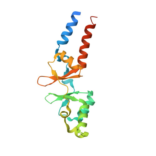
    6ZZ9, 6ZZA

  • PubMed Abstract: 

    Cyclic di-AMP is an essential signalling molecule in Gram-positive bacteria. This second messenger regulates the osmotic pressure of the cell by interacting directly with the regulatory domains, either RCK_C or CBS domains, of several potassium and osmolyte uptake membrane protein systems. Cyclic di-AMP also targets stand-alone CBS domain proteins such as DarB in Bacillus subtilis and CbpB in Listeria monocytogenes. We show here that the CbpB protein of Group B Streptococcus binds c-di-AMP with a very high affinity. Crystal structures of CbpB reveal the determinants of binding specificity and significant conformational changes occurring upon c-di-AMP binding. Deletion of the cbpB gene alters bacterial growth in low potassium conditions most likely due to a decrease in the amount of ppGpp caused by a loss of interaction between CbpB and Rel, the GTP/GDP pyrophosphokinase.


  • Organizational Affiliation
    • Unité Biologie des Bactéries Pathogènes à Gram-positif, CNRS UMR 6047, Institut Pasteur, Université Paris Cité, France.

Macromolecule Content 

  • Total Structure Weight: 20.6 kDa 
  • Atom Count: 1,288 
  • Modeled Residue Count: 150 
  • Deposited Residue Count: 176 
  • Unique protein chains: 1

Macromolecules

Find similar proteins by:|  3D Structure
Entity ID: 1
MoleculeChains  Sequence LengthOrganismDetailsImage
CBS domain-containing protein176Streptococcus agalactiaeMutation(s): 0 
Gene Names: ykuLC6N06_01440D5F95_08070DX05_08405E8E04_03435F5F86_02645NCTC9828_00932WA02_02795WA05_04310
UniProt
Find proteins for A0A076YWK5 (Streptococcus agalactiae)
Explore A0A076YWK5 
Go to UniProtKB:  A0A076YWK5
Entity Groups
Sequence Clusters30% Identity50% Identity70% Identity90% Identity95% Identity100% Identity
UniProt GroupA0A076YWK5
Sequence Annotations
Expand
Reference Sequence

Small Molecules

Ligands 2 Unique
IDChains Name / Formula / InChI Key2D Diagram3D Interactions
2BA
(Subject of Investigation/LOI)

Query on 2BA


Download:Ideal Coordinates CCD File
B [auth A](2R,3R,3aS,5R,7aR,9R,10R,10aS,12R,14aR)-2,9-bis(6-amino-9H-purin-9-yl)octahydro-2H,7H-difuro[3,2-d:3',2'-j][1,3,7,9,2,8 ]tetraoxadiphosphacyclododecine-3,5,10,12-tetrol 5,12-dioxide
C20 H24 N10 O12 P2
PDXMFTWFFKBFIN-XPWFQUROSA-N
SO4

Query on SO4


Download:Ideal Coordinates CCD File
C [auth A]SULFATE ION
O4 S
QAOWNCQODCNURD-UHFFFAOYSA-L

Experimental Data & Validation

Experimental Data

  • Method: X-RAY DIFFRACTION
  • Resolution: 2.49 Å
  • R-Value Free:  0.245 (Depositor), 0.259 (DCC) 
  • R-Value Work:  0.186 (Depositor), 0.193 (DCC) 
  • R-Value Observed: 0.189 (Depositor) 
Space Group: P 31 2 1
Unit Cell:
Length ( Å )Angle ( ˚ )
a = 93.83α = 90
b = 93.83β = 90
c = 33.6γ = 120
Software Package:
Software NamePurpose
BUSTERrefinement
PHASERphasing
Aimlessdata scaling
XDSdata reduction

Structure Validation

View Full Validation Report


Ligand Structure Quality Assessment 


Entry History 

Deposition Data

Revision History  (Full details and data files)

  • Version 1.0: 2021-08-18
    Type: Initial release
  • Version 1.1: 2024-01-31
    Changes: Data collection, Refinement description
  • Version 1.2: 2025-08-13
    Changes: Database references, Structure summary