6ZT1 | pdb_00006zt1

Two states of a de novo hexameric coiled coil assembly CC-Type2-(LaIdGe)4


Experimental Data Snapshot

  • Method: X-RAY DIFFRACTION
  • Resolution: 2.01 Å
  • R-Value Free: 
    0.261 (Depositor), 0.259 (DCC) 
  • R-Value Work: 
    0.207 (Depositor) 
  • R-Value Observed: 
    0.210 (Depositor) 

wwPDB Validation 3D Report Full Report

Validation slider image for 6ZT1

This is version 1.2 of the entry. See complete history

Literature

Structural resolution of switchable states of a de novo peptide assembly.

Dawson, W.M.Lang, E.J.M.Rhys, G.G.Shelley, K.L.Williams, C.Brady, R.L.Crump, M.P.Mulholland, A.J.Woolfson, D.N.

(2021) Nat Commun 12: 1530-1530

  • DOI: https://doi.org/10.1038/s41467-021-21851-8
  • Primary Citation Related Structures: 
    6ZT1

  • PubMed Abstract: 

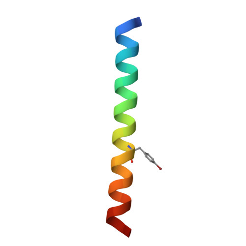
    De novo protein design is advancing rapidly. However, most designs are for single states. Here we report a de novo designed peptide that forms multiple α-helical-bundle states that are accessible and interconvertible under the same conditions. Usually in such designs amphipathic α helices associate to form compact structures with consolidated hydrophobic cores. However, recent rational and computational designs have delivered open α-helical barrels with functionalisable cavities. By placing glycine judiciously in the helical interfaces of an α-helical barrel, we obtain both open and compact states in a single protein crystal. Molecular dynamics simulations indicate a free-energy landscape with multiple and interconverting states. Together, these findings suggest a frustrated system in which steric interactions that maintain the open barrel and the hydrophobic effect that drives complete collapse are traded-off. Indeed, addition of a hydrophobic co-solvent that can bind within the barrel affects the switch between the states both in silico and experimentally.


  • Organizational Affiliation
    • School of Chemistry, University of Bristol, Cantock's Close, Bristol, UK.

Macromolecule Content 

  • Total Structure Weight: 38.62 kDa 
  • Atom Count: 2,806 
  • Modeled Residue Count: 364 
  • Deposited Residue Count: 384 
  • Unique protein chains: 1

Macromolecules

Find similar proteins by:|  3D Structure
Entity ID: 1
MoleculeChains  Sequence LengthOrganismDetailsImage
CC-Type2-(LaIdGe)4
A, B, C, D, E
A, B, C, D, E, F, G, H, I, J, K, L
32synthetic constructMutation(s): 0 

Small Molecules

Modified Residues  1 Unique
IDChains TypeFormula2D DiagramParent
4BF
Query on 4BF
A, B, C, D, E
A, B, C, D, E, F, G, H, I, J, K, L
L-PEPTIDE LINKINGC9 H10 Br N O2TYR

Experimental Data & Validation

Experimental Data

  • Method: X-RAY DIFFRACTION
  • Resolution: 2.01 Å
  • R-Value Free:  0.261 (Depositor), 0.259 (DCC) 
  • R-Value Work:  0.207 (Depositor) 
  • R-Value Observed: 0.210 (Depositor) 
Space Group: P 21 21 2
Unit Cell:
Length ( Å )Angle ( ˚ )
a = 58.92α = 90
b = 128.1β = 90
c = 56.53γ = 90
Software Package:
Software NamePurpose
PHENIXrefinement
PDB_EXTRACTdata extraction
xia2data reduction
xia2data scaling
CRANK2phasing

Structure Validation

View Full Validation Report


Entry History 

& Funding Information

Deposition Data


Funding OrganizationLocationGrant Number
European Research Council (ERC)European Union340764

Revision History  (Full details and data files)

  • Version 1.0: 2021-02-03
    Type: Initial release
  • Version 1.1: 2021-04-07
    Changes: Database references
  • Version 1.2: 2025-04-09
    Changes: Advisory, Data collection, Database references, Refinement description, Structure summary