6ZP5 | pdb_00006zp5

SARS-CoV-2 spike in prefusion state (flexibility analysis, 1-up closed conformation)


Experimental Data Snapshot

  • Method: ELECTRON MICROSCOPY
  • Resolution: 3.10 Å
  • Aggregation State: PARTICLE 
  • Reconstruction Method: SINGLE PARTICLE 

Starting Model: experimental
View more details

wwPDB Validation 3D Report Full Report

Validation slider image for 6ZP5

This is version 2.2 of the entry. See complete history

Literature

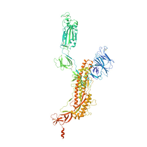
Continuous flexibility analysis of SARS-CoV-2 spike prefusion structures.

Melero, R.Sorzano, C.O.S.Foster, B.Vilas, J.L.Martinez, M.Marabini, R.Ramirez-Aportela, E.Sanchez-Garcia, R.Herreros, D.Del Cano, L.Losana, P.Fonseca-Reyna, Y.C.Conesa, P.Wrapp, D.Chacon, P.McLellan, J.S.Tagare, H.D.Carazo, J.M.

(2020) IUCrJ 7: 1059-1069

  • DOI: https://doi.org/10.1107/S2052252520012725
  • Primary Citation Related Structures: 
    6ZOW, 6ZP5, 6ZP7

  • PubMed Abstract: 

    Using a new consensus-based image-processing approach together with principal component analysis, the flexibility and conformational dynamics of the SARS-CoV-2 spike in the prefusion state have been analysed. These studies revealed concerted motions involving the receptor-binding domain (RBD), N-terminal domain, and subdomains 1 and 2 around the previously characterized 1-RBD-up state, which have been modeled as elastic deformations. It is shown that in this data set there are not well defined, stable spike conformations, but virtually a continuum of states. An ensemble map was obtained with minimum bias, from which the extremes of the change along the direction of maximal variance were modeled by flexible fitting. The results provide a warning of the potential image-processing classification instability of these complicated data sets, which has a direct impact on the interpretability of the results.


  • Organizational Affiliation
    • Centro Nacional de Biotecnologia-CSIC, Calle Darwin 3, 28049 Cantoblanco, Madrid, Spain.

Macromolecule Content 

  • Total Structure Weight: 445.67 kDa 
  • Atom Count: 23,615 
  • Modeled Residue Count: 2,861 
  • Deposited Residue Count: 3,864 
  • Unique protein chains: 1

Macromolecules

Find similar proteins by:|  3D Structure
Entity ID: 1
MoleculeChains  Sequence LengthOrganismDetailsImage
Spike glycoprotein
A, B, C
1,288Severe acute respiratory syndrome coronavirus 2Mutation(s): 5 
Gene Names: S2
UniProt
Find proteins for P0DTC2 (Severe acute respiratory syndrome coronavirus 2)
Explore P0DTC2 
Go to UniProtKB:  P0DTC2
Entity Groups
Sequence Clusters30% Identity50% Identity70% Identity90% Identity95% Identity100% Identity
UniProt GroupP0DTC2
Glycosylation
Glycosylation Sites: 3Go to GlyGen: P0DTC2-1
Sequence Annotations
Expand
Reference Sequence

Oligosaccharides

Help  
Entity ID: 3
MoleculeChains Length2D Diagram GlycosylationD Interactions
alpha-D-mannopyranose-(1-3)-beta-D-mannopyranose-(1-4)-2-acetamido-2-deoxy-beta-D-glucopyranose-(1-4)-2-acetamido-2-deoxy-beta-D-glucopyranose
E, F, R, U
4N-Glycosylation
Glycosylation Resources
GlyTouCan: G81315DD
GlyCosmos: G81315DD
GlyGen: G81315DD
Entity ID: 4
MoleculeChains Length2D Diagram GlycosylationD Interactions
beta-D-mannopyranose-(1-4)-2-acetamido-2-deoxy-beta-D-glucopyranose-(1-4)-2-acetamido-2-deoxy-beta-D-glucopyranose
AA [auth a],
BA [auth b],
N,
O,
W,
AA [auth a],
BA [auth b],
N,
O,
W,
Y
3N-Glycosylation
Glycosylation Resources
GlyTouCan: G15407YE
GlyCosmos: G15407YE
GlyGen: G15407YE
Entity ID: 5
MoleculeChains Length2D Diagram GlycosylationD Interactions
beta-D-mannopyranose-(1-4)-2-acetamido-2-deoxy-beta-D-glucopyranose-(1-6)-2-acetamido-2-deoxy-beta-D-glucopyranose
Q
3N/A
Glycosylation Resources
GlyTouCan: G62916OK
GlyCosmos: G62916OK
GlyGen: G62916OK

Small Molecules

Ligands 3 Unique
IDChains Name / Formula / InChI Key2D Diagram3D Interactions
NAG

Query on NAG


Download:Ideal Coordinates CCD File
AB [auth C]
EA [auth A]
FA [auth A]
GA [auth A]
HA [auth A]
AB [auth C],
EA [auth A],
FA [auth A],
GA [auth A],
HA [auth A],
IA [auth A],
JA [auth A],
OA [auth B],
PA [auth B],
QA [auth B],
RA [auth B],
SA [auth B],
TA [auth B],
UA [auth B],
VA [auth B],
WA [auth C],
XA [auth C],
YA [auth C]
2-acetamido-2-deoxy-beta-D-glucopyranose
C8 H15 N O6
OVRNDRQMDRJTHS-FMDGEEDCSA-N
MAN

Query on MAN


Download:Ideal Coordinates CCD File
KA [auth A],
ZA [auth C]
alpha-D-mannopyranose
C6 H12 O6
WQZGKKKJIJFFOK-PQMKYFCFSA-N
DMS

Query on DMS


Download:Ideal Coordinates CCD File
LA [auth A],
MA [auth A],
NA [auth A]
DIMETHYL SULFOXIDE
C2 H6 O S
IAZDPXIOMUYVGZ-UHFFFAOYSA-N

Experimental Data & Validation

Experimental Data

  • Method: ELECTRON MICROSCOPY
  • Resolution: 3.10 Å
  • Aggregation State: PARTICLE 
  • Reconstruction Method: SINGLE PARTICLE 

Structure Validation

View Full Validation Report


Entry History 

& Funding Information

Deposition Data


Funding OrganizationLocationGrant Number
Spanish National Research CouncilSpainPIE/COVID-19 202020E079
Spanish Ministry of Science, Innovation, and UniversitiesSpainSEV 2017-0712
European Research Council (ERC)SpainERC - 2018 - SyG, Proposal: 810057
National Institutes of Health/National Library of Medicine (NIH/NLM)United StatesGM125769
National Institutes of Health/National Library of Medicine (NIH/NLM)United StatesR01-AI127521

Revision History  (Full details and data files)

  • Version 1.0: 2020-07-29
    Type: Initial release
  • Version 2.0: 2021-02-03
    Changes: Advisory, Atomic model, Data collection, Database references, Derived calculations, Polymer sequence, Source and taxonomy, Structure summary
  • Version 2.1: 2022-12-21
    Changes: Database references, Refinement description
  • Version 2.2: 2024-10-23
    Changes: Data collection, Refinement description, Structure summary