6ZNS | pdb_00006zns

Crystal Structure of DUF1998 helicase MrfA


Experimental Data Snapshot

  • Method: X-RAY DIFFRACTION
  • Resolution: 3.32 Å
  • R-Value Free: 
    0.342 (Depositor), 0.346 (DCC) 
  • R-Value Work: 
    0.307 (Depositor), 0.314 (DCC) 
  • R-Value Observed: 
    0.309 (Depositor) 

Starting Model: experimental
View more details

wwPDB Validation 3D Report Full Report

Validation slider image for 6ZNS

This is version 1.5 of the entry. See complete history

Literature

A skipping rope translocation mechanism in a widespread family of DNA repair helicases.

Roske, J.J.Liu, S.Loll, B.Neu, U.Wahl, M.C.

(2021) Nucleic Acids Res 49: 504-518

  • DOI: https://doi.org/10.1093/nar/gkaa1174
  • Primary Citation Related Structures: 
    6ZNP, 6ZNQ, 6ZNS

  • PubMed Abstract: 

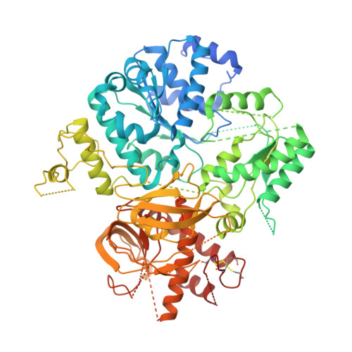
    Mitomycin repair factor A represents a family of DNA helicases that harbor a domain of unknown function (DUF1998) and support repair of mitomycin C-induced DNA damage by presently unknown molecular mechanisms. We determined crystal structures of Bacillus subtilis Mitomycin repair factor A alone and in complex with an ATP analog and/or DNA and conducted structure-informed functional analyses. Our results reveal a unique set of auxiliary domains appended to a dual-RecA domain core. Upon DNA binding, a Zn2+-binding domain, encompassing the domain of unknown function, acts like a drum that rolls out a canopy of helicase-associated domains, entrapping the substrate and tautening an inter-domain linker across the loading strand. Quantification of DNA binding, stimulated ATPase and helicase activities in the wild type and mutant enzyme variants in conjunction with the mode of coordination of the ATP analog suggest that Mitomycin repair factor A employs similar ATPase-driven conformational changes to translocate on DNA, with the linker ratcheting through the nucleotides like a 'skipping rope'. The electrostatic surface topology outlines a likely path for the displaced DNA strand. Our results reveal unique molecular mechanisms in a widespread family of DNA repair helicases linked to bacterial antibiotics resistance.


  • Organizational Affiliation
    • Freie Universität Berlin, Institute of Chemistry and Biochemistry, Laboratory of Structural Biochemistry, Takustraβe 6, D-14195 Berlin, Germany.

Macromolecule Content 

  • Total Structure Weight: 84.9 kDa 
  • Atom Count: 5,057 
  • Modeled Residue Count: 635 
  • Deposited Residue Count: 751 
  • Unique protein chains: 1

Macromolecules

Find similar proteins by:|  3D Structure
Entity ID: 1
MoleculeChains  Sequence LengthOrganismDetailsImage
Uncharacterized ATP-dependent helicase YprA751Bacillus subtilis subsp. subtilis str. 168Mutation(s): 0 
Gene Names: yprABSU22220
EC: 3.6.4
UniProt
Find proteins for P50830 (Bacillus subtilis (strain 168))
Explore P50830 
Go to UniProtKB:  P50830
Entity Groups
Sequence Clusters30% Identity50% Identity70% Identity90% Identity95% Identity100% Identity
UniProt GroupP50830
Sequence Annotations
Expand
Reference Sequence

Small Molecules

Ligands 1 Unique
IDChains Name / Formula / InChI Key2D Diagram3D Interactions
ZN

Query on ZN


Download:Ideal Coordinates CCD File
B [auth A]ZINC ION
Zn
PTFCDOFLOPIGGS-UHFFFAOYSA-N

Experimental Data & Validation

Experimental Data

  • Method: X-RAY DIFFRACTION
  • Resolution: 3.32 Å
  • R-Value Free:  0.342 (Depositor), 0.346 (DCC) 
  • R-Value Work:  0.307 (Depositor), 0.314 (DCC) 
  • R-Value Observed: 0.309 (Depositor) 
Space Group: H 3
Unit Cell:
Length ( Å )Angle ( ˚ )
a = 191.943α = 90
b = 191.943β = 90
c = 65.892γ = 120
Software Package:
Software NamePurpose
PHENIXrefinement
Cootmodel building
MxCuBEdata collection
PHASERphasing
XDSdata reduction
XSCALEdata scaling

Structure Validation

View Full Validation Report


Entry History 

& Funding Information

Deposition Data


Funding OrganizationLocationGrant Number
German Research Foundation (DFG)GermanyRTG 2473-1

Revision History  (Full details and data files)

  • Version 1.0: 2020-11-25
    Type: Initial release
  • Version 1.1: 2020-12-16
    Changes: Database references
  • Version 1.2: 2020-12-23
    Changes: Database references
  • Version 1.3: 2021-01-20
    Changes: Database references
  • Version 1.4: 2024-05-01
    Changes: Data collection, Database references, Refinement description
  • Version 1.5: 2024-11-13
    Changes: Structure summary