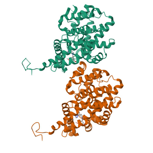
6ZND

[1,2,4]Triazolo[1,5-a]pyrimidine Phosphodiesterase 2 Inhibitors


Experimental Data Snapshot

  • Method: X-RAY DIFFRACTION
  • Resolution: 2.35 Å
  • R-Value Free: 0.291 
  • R-Value Work: 0.215 
  • R-Value Observed: 0.218 

Starting Model: experimental
View more details

wwPDB Validation   3D Report Full Report


Ligand Structure Quality Assessment 


This is version 1.3 of the entry. See complete history


Literature

[1,2,4]Triazolo[1,5- a ]pyrimidine Phosphodiesterase 2A Inhibitors: Structure and Free-Energy Perturbation-Guided Exploration.

Tresadern, G.Velter, I.Trabanco, A.A.Van den Keybus, F.Macdonald, G.J.Somers, M.V.F.Vanhoof, G.Leonard, P.M.Lamers, M.B.A.C.Van Roosbroeck, Y.E.M.Buijnsters, P.J.J.A.

(2020) J Med Chem 63: 12887-12910

  • DOI: https://doi.org/10.1021/acs.jmedchem.0c01272
  • Primary Citation of Related Structures:  
    6ZND, 6ZQZ

  • PubMed Abstract: 

    We describe the hit-to-lead exploration of a [1,2,4]triazolo[1,5- a ]pyrimidine phosphodiesterase 2A (PDE2A) inhibitor arising from high-throughput screening. X-ray crystallography enabled structure-guided design, leading to the identification of preferred substructural components. Further rounds of optimization used relative binding free-energy calculations to prioritize different substituents from the large accessible chemical space. The free-energy perturbation (FEP) calculations were performed for 265 putative PDE2A inhibitors, and 100 compounds were synthesized representing a relatively large prospective application providing unexpectedly active molecules with IC 50 's from 2340 to 0.89 nM. Lead compound 46 originating from the FEP calculations showed PDE2A inhibition IC 50 of 1.3 ± 0.39 nM, ∼100-fold selectivity versus other PDE enzymes, clean cytochrome P450 profile, in vivo target occupancy, and promise for further lead optimization.


  • Organizational Affiliation

    Computational Chemistry, Janssen Pharmaceutica N. V., Turnhoutseweg 30, B-2340 Beerse, Belgium.


Macromolecules
Find similar proteins by:  (by identity cutoff)  |  3D Structure
Entity ID: 1
MoleculeChains Sequence LengthOrganismDetailsImage
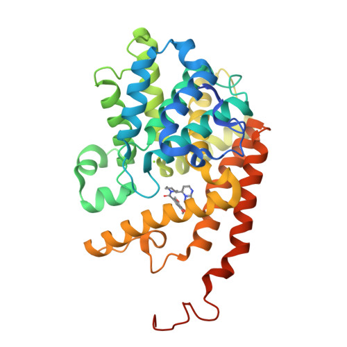
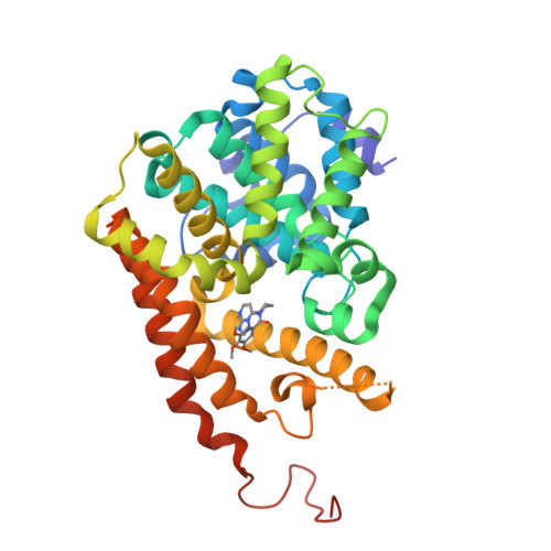
cGMP-dependent 3',5'-cyclic phosphodiesterase
A, B
353Homo sapiensMutation(s): 0 
Gene Names: PDE2A
EC: 3.1.4.17
UniProt & NIH Common Fund Data Resources
Find proteins for O00408 (Homo sapiens)
Explore O00408 
Go to UniProtKB:  O00408
PHAROS:  O00408
GTEx:  ENSG00000186642 
Entity Groups  
Sequence Clusters30% Identity50% Identity70% Identity90% Identity95% Identity100% Identity
UniProt GroupO00408
Sequence Annotations
Expand
  • Reference Sequence
Small Molecules
Ligands 3 Unique
IDChains Name / Formula / InChI Key2D Diagram3D Interactions
QMZ (Subject of Investigation/LOI)
Query on QMZ

Download Ideal Coordinates CCD File 
E [auth A],
H [auth B]
[(3~{S})-3-([1,2,4]triazolo[1,5-a]pyrimidin-7-yl)piperidin-1-yl]-(3,4,5-trimethoxyphenyl)methanone
C20 H23 N5 O4
QRHYHQMFAMRYKQ-ZDUSSCGKSA-N
ZN
Query on ZN

Download Ideal Coordinates CCD File 
C [auth A],
F [auth B]
ZINC ION
Zn
PTFCDOFLOPIGGS-UHFFFAOYSA-N
MG
Query on MG

Download Ideal Coordinates CCD File 
D [auth A],
G [auth B]
MAGNESIUM ION
Mg
JLVVSXFLKOJNIY-UHFFFAOYSA-N
Experimental Data & Validation

Experimental Data

  • Method: X-RAY DIFFRACTION
  • Resolution: 2.35 Å
  • R-Value Free: 0.291 
  • R-Value Work: 0.215 
  • R-Value Observed: 0.218 
  • Space Group: C 1 2 1
Unit Cell:
Length ( Å )Angle ( ˚ )
a = 171.06α = 90
b = 69.52β = 91.16
c = 55.09γ = 90
Software Package:
Software NamePurpose
REFMACrefinement
PDB_EXTRACTdata extraction
iMOSFLMdata reduction
SCALAdata scaling
PHASERphasing

Structure Validation

View Full Validation Report


Ligand Structure Quality Assessment 


Entry History 

Deposition Data

Revision History  (Full details and data files)

  • Version 1.0: 2020-07-22
    Type: Initial release
  • Version 1.1: 2020-11-18
    Changes: Database references, Derived calculations
  • Version 1.2: 2020-11-25
    Changes: Database references
  • Version 1.3: 2024-01-31
    Changes: Data collection, Database references, Refinement description