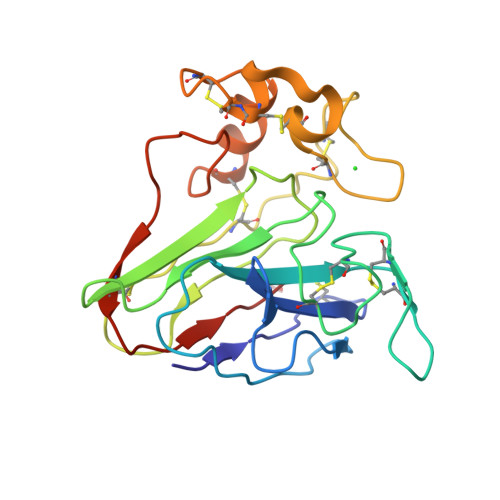
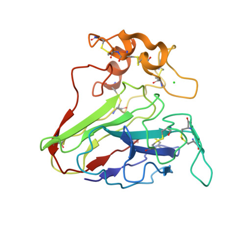
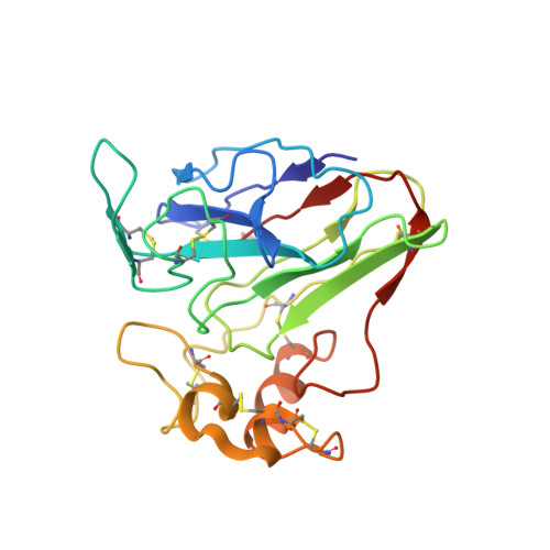
6ZHN

3D electron diffraction structure of thaumatin from Thaumatococcus daniellii


Experimental Data Snapshot

  • Method: ELECTRON CRYSTALLOGRAPHY
  • Resolution: 2.76 Å
  • R-Value Free: 0.321 
  • R-Value Work: 0.280 
  • R-Value Observed: 0.282 

Starting Model: experimental
View more details

wwPDB Validation   3D Report Full Report


This is version 1.3 of the entry. See complete history


Literature

Statistically correcting dynamical electron scattering improves the refinement of protein nanocrystals, including charge refinement of coordinated metals.

Blum, T.B.Housset, D.Clabbers, M.T.B.van Genderen, E.Bacia-Verloop, M.Zander, U.McCarthy, A.A.Schoehn, G.Ling, W.L.Abrahams, J.P.

(2021) Acta Crystallogr D Struct Biol 77: 75-85

  • DOI: https://doi.org/10.1107/S2059798320014540
  • Primary Citation of Related Structures:  
    6ZHB, 6ZHJ, 6ZHN, 6ZI8

  • PubMed Abstract: 

    Electron diffraction allows protein structure determination when only nanosized crystals are available. Nevertheless, multiple elastic (or dynamical) scattering, which is prominent in electron diffraction, is a concern. Current methods for modeling dynamical scattering by multi-slice or Bloch wave approaches are not suitable for protein crystals because they are not designed to cope with large molecules. Here, dynamical scattering of nanocrystals of insulin, thermolysin and thaumatin was limited by collecting data from thin crystals. To accurately measure the weak diffraction signal from the few unit cells in the thin crystals, a low-noise hybrid pixel Timepix electron-counting detector was used. The remaining dynamical component was further reduced in refinement using a likelihood-based correction, which was introduced previously for analyzing electron diffraction data of small-molecule nanocrystals and was adapted here for protein crystals. The procedure is shown to notably improve the structural refinement, in one case allowing the location of solvent molecules. It also allowed refinement of the charge states of bound metal atoms, an important element in protein function, through B-factor analysis of the metal atoms and their ligands. These results clearly increase the value of macromolecular electron crystallography as a complementary structural biology technique.


  • Organizational Affiliation

    Department of Biology and Chemistry, Paul Scherrer Institute, 5232 Villigen, Switzerland.


Macromolecules
Find similar proteins by:  (by identity cutoff)  |  3D Structure
Entity ID: 1
MoleculeChains Sequence LengthOrganismDetailsImage
Thaumatin-1207Thaumatococcus danielliiMutation(s): 0 
UniProt
Find proteins for P02883 (Thaumatococcus daniellii)
Explore P02883 
Go to UniProtKB:  P02883
Entity Groups  
Sequence Clusters30% Identity50% Identity70% Identity90% Identity95% Identity100% Identity
UniProt GroupP02883
Sequence Annotations
Expand
  • Reference Sequence
Small Molecules
Ligands 1 Unique
IDChains Name / Formula / InChI Key2D Diagram3D Interactions
CL
Query on CL

Download Ideal Coordinates CCD File 
B [auth A]CHLORIDE ION
Cl
VEXZGXHMUGYJMC-UHFFFAOYSA-M
Experimental Data & Validation

Experimental Data

  • Method: ELECTRON CRYSTALLOGRAPHY
  • Resolution: 2.76 Å
  • R-Value Free: 0.321 
  • R-Value Work: 0.280 
  • R-Value Observed: 0.282 
  • Space Group: P 41 21 2
Unit Cell:
Length ( Å )Angle ( ˚ )
a = 57.72α = 90
b = 57.72β = 90
c = 149.17γ = 90
Software Package:
Software NamePurpose
REFMACrefinement
PDB_EXTRACTdata extraction
PHASERphasing

Structure Validation

View Full Validation Report


Entry History & Funding Information

Deposition Data


Funding OrganizationLocationGrant Number
French National Research AgencyFranceANR-10-INSB-05-02
French National Research AgencyFranceANR-10-LABX-49-01
Swiss National Science FoundationSwitzerland31003A_17002
Swiss National Science FoundationSwitzerland200021_165669

Revision History  (Full details and data files)

  • Version 1.0: 2021-01-27
    Type: Initial release
  • Version 1.1: 2023-09-13
    Changes: Data collection, Database references
  • Version 1.2: 2024-01-24
    Changes: Refinement description
  • Version 1.3: 2024-10-16
    Changes: Structure summary