6ZGF | pdb_00006zgf

Spike Protein of RaTG13 Bat Coronavirus in Closed Conformation


Experimental Data Snapshot

  • Method: ELECTRON MICROSCOPY
  • Resolution: 3.10 Å
  • Aggregation State: PARTICLE 
  • Reconstruction Method: SINGLE PARTICLE 

Starting Model: experimental
View more details

wwPDB Validation 3D Report Full Report

Validation slider image for 6ZGF

This is version 2.2 of the entry. See complete history

Literature

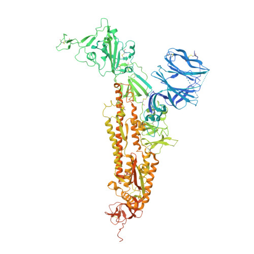
SARS-CoV-2 and bat RaTG13 spike glycoprotein structures inform on virus evolution and furin-cleavage effects.

Wrobel, A.G.Benton, D.J.Xu, P.Roustan, C.Martin, S.R.Rosenthal, P.B.Skehel, J.J.Gamblin, S.J.

(2020) Nat Struct Mol Biol 27: 763-767

  • DOI: https://doi.org/10.1038/s41594-020-0468-7
  • Primary Citation Related Structures: 
    6ZGE, 6ZGF, 6ZGG, 6ZGH, 6ZGI

  • PubMed Abstract: 

    SARS-CoV-2 is thought to have emerged from bats, possibly via a secondary host. Here, we investigate the relationship of spike (S) glycoprotein from SARS-CoV-2 with the S protein of a closely related bat virus, RaTG13. We determined cryo-EM structures for RaTG13 S and for both furin-cleaved and uncleaved SARS-CoV-2 S; we compared these with recently reported structures for uncleaved SARS-CoV-2 S. We also biochemically characterized their relative stabilities and affinities for the SARS-CoV-2 receptor ACE2. Although the overall structures of human and bat virus S proteins are similar, there are key differences in their properties, including a more stable precleavage form of human S and about 1,000-fold tighter binding of SARS-CoV-2 to human receptor. These observations suggest that cleavage at the furin-cleavage site decreases the overall stability of SARS-CoV-2 S and facilitates the adoption of the open conformation that is required for S to bind to the ACE2 receptor.


  • Organizational Affiliation
    • Structural Biology of Disease Processes Laboratory, Francis Crick Institute, London, UK. antoni.wrobel@crick.ac.uk.

Macromolecule Content 

  • Total Structure Weight: 445.09 kDa 
  • Atom Count: 26,163 
  • Modeled Residue Count: 3,180 
  • Deposited Residue Count: 3,849 
  • Unique protein chains: 1

Macromolecules

Find similar proteins by:|  3D Structure
Entity ID: 1
MoleculeChains  Sequence LengthOrganismDetailsImage
Spike glycoprotein
A, B, C
1,283Bat coronavirus RaTG13Mutation(s): 0 

Oligosaccharides

Help  
Entity ID: 3
MoleculeChains Length2D Diagram GlycosylationD Interactions
2-acetamido-2-deoxy-beta-D-glucopyranose-(1-4)-[alpha-L-fucopyranose-(1-6)]2-acetamido-2-deoxy-beta-D-glucopyranoseBA [auth b],
H,
R
3N-Glycosylation
Glycosylation Resources
GlyTouCan: G21290RB
GlyCosmos: G21290RB
GlyGen: G21290RB
Entity ID: 4
MoleculeChains Length2D Diagram GlycosylationD Interactions
beta-D-mannopyranose-(1-4)-2-acetamido-2-deoxy-beta-D-glucopyranose-(1-4)-2-acetamido-2-deoxy-beta-D-glucopyranoseCA [auth c],
I,
S
3N-Glycosylation
Glycosylation Resources
GlyTouCan: G15407YE
GlyCosmos: G15407YE
GlyGen: G15407YE
Entity ID: 5
MoleculeChains Length2D Diagram GlycosylationD Interactions
alpha-L-fucopyranose-(1-6)-2-acetamido-2-deoxy-beta-D-glucopyranoseGA [auth g],
M,
W
2N-Glycosylation
Glycosylation Resources
GlyTouCan: G86851RC
GlyCosmos: G86851RC
GlyGen: G86851RC

Small Molecules

Ligands 1 Unique
IDChains Name / Formula / InChI Key2D Diagram3D Interactions
NAG

Query on NAG


Download:Ideal Coordinates CCD File
AB [auth C]
BB [auth C]
CB [auth C]
DB [auth C]
EB [auth C]
AB [auth C],
BB [auth C],
CB [auth C],
DB [auth C],
EB [auth C],
FB [auth C],
GB [auth C],
HA [auth A],
HB [auth C],
IA [auth A],
JA [auth A],
KA [auth A],
LA [auth A],
MA [auth A],
NA [auth A],
OA [auth A],
PA [auth A],
QA [auth B],
RA [auth B],
SA [auth B],
TA [auth B],
UA [auth B],
VA [auth B],
WA [auth B],
XA [auth B],
YA [auth B],
ZA [auth C]
2-acetamido-2-deoxy-beta-D-glucopyranose
C8 H15 N O6
OVRNDRQMDRJTHS-FMDGEEDCSA-N

Experimental Data & Validation

Experimental Data

  • Method: ELECTRON MICROSCOPY
  • Resolution: 3.10 Å
  • Aggregation State: PARTICLE 
  • Reconstruction Method: SINGLE PARTICLE 
EM Software:
TaskSoftware PackageVersion
MODEL REFINEMENTPHENIX1.18
RECONSTRUCTIONcryoSPARC2.15

Structure Validation

View Full Validation Report


Entry History 

& Funding Information

Deposition Data


Funding OrganizationLocationGrant Number
The Francis Crick InstituteUnited KingdomFC001078
The Francis Crick InstituteUnited KingdomFC001143

Revision History  (Full details and data files)

  • Version 1.0: 2020-07-01
    Type: Initial release
  • Version 2.0: 2020-07-29
    Type: Remediation
    Reason: Carbohydrate remediation
    Changes: Atomic model, Data collection, Derived calculations, Structure summary
  • Version 2.1: 2020-09-16
    Changes: Database references, Structure summary
  • Version 2.2: 2024-11-13
    Changes: Data collection, Database references, Refinement description, Structure summary