6ZEV | pdb_00006zev

Crystal structure of proteinase K lamellae by electron diffraction with a 20 micrometre C2 condenser aperture


Experimental Data Snapshot

  • Method: ELECTRON CRYSTALLOGRAPHY
  • Resolution: 2.40 Å
  • R-Value Free: 
    0.243 (Depositor), 0.255 (DCC) 
  • R-Value Work: 
    0.200 (Depositor), 0.215 (DCC) 

Starting Model: experimental
View more details

wwPDB Validation 3D Report Full Report

Validation slider image for 6ZEV

This is version 1.5 of the entry. See complete history

Literature

A Workflow for Protein Structure Determination From Thin Crystal Lamella by Micro-Electron Diffraction.

Beale, E.V.Waterman, D.G.Hecksel, C.van Rooyen, J.Gilchrist, J.B.Parkhurst, J.M.de Haas, F.Buijsse, B.Evans, G.Zhang, P.

(2020) Front Mol Biosci 7: 179-179

  • DOI: https://doi.org/10.3389/fmolb.2020.00179
  • Primary Citation Related Structures: 
    6ZET, 6ZEU, 6ZEV

  • PubMed Abstract: 

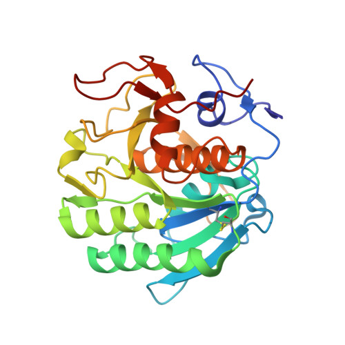
    MicroED has recently emerged as a powerful method for the analysis of biological structures at atomic resolution. This technique has been largely limited to protein nanocrystals which grow either as needles or plates measuring only a few hundred nanometers in thickness. Furthermore, traditional microED data processing uses established X-ray crystallography software that is not optimized for handling compound effects that are unique to electron diffraction data. Here, we present an integrated workflow for microED, from sample preparation by cryo-focused ion beam milling, through data collection with a standard Ceta-D detector, to data processing using the DIALS software suite, thus enabling routine atomic structure determination of protein crystals of any size and shape using microED. We demonstrate the effectiveness of the workflow by determining the structure of proteinase K to 2.0 Å resolution and show the advantage of using protein crystal lamellae over nanocrystals.


  • Organizational Affiliation
    • Diamond Light Source, Harwell Science and Innovation Campus, Didcot, United Kingdom.

Macromolecule Content 

  • Total Structure Weight: 29 kDa 
  • Atom Count: 2,060 
  • Modeled Residue Count: 278 
  • Deposited Residue Count: 279 
  • Unique protein chains: 1

Macromolecules

Find similar proteins by:|  3D Structure
Entity ID: 1
MoleculeChains  Sequence LengthOrganismDetailsImage
Proteinase KA [auth AAA]279Parengyodontium albumMutation(s): 0 
EC: 3.4.21.64
UniProt
Find proteins for P06873 (Parengyodontium album)
Explore P06873 
Go to UniProtKB:  P06873
Entity Groups
Sequence Clusters30% Identity50% Identity70% Identity90% Identity95% Identity100% Identity
UniProt GroupP06873
Sequence Annotations
Expand
Reference Sequence

Small Molecules

Ligands 1 Unique
IDChains Name / Formula / InChI Key2D Diagram3D Interactions
CA

Query on CA


Download:Ideal Coordinates CCD File
B [auth AAA]CALCIUM ION
Ca
BHPQYMZQTOCNFJ-UHFFFAOYSA-N

Experimental Data & Validation

Experimental Data

  • Method: ELECTRON CRYSTALLOGRAPHY
  • Resolution: 2.40 Å
  • R-Value Free:  0.243 (Depositor), 0.255 (DCC) 
  • R-Value Work:  0.200 (Depositor), 0.215 (DCC) 
Space Group: P 43 21 2
Unit Cell:
Length ( Å )Angle ( ˚ )
a = 67.328α = 90
b = 67.328β = 90
c = 106.599γ = 90
Software Package:
Software NamePurpose
REFMACrefinement
DIALSdata reduction
Aimlessdata scaling
PHASERphasing
EM Software:
TaskSoftware PackageVersion
MODEL REFINEMENTREFMAC5.8.0258

Structure Validation

View Full Validation Report


Entry History 

& Funding Information

Deposition Data


Funding OrganizationLocationGrant Number
Wellcome TrustUnited Kingdom206422/Z/17/Z
Biotechnology and Biological Sciences Research Council (BBSRC)United KingdomBB/S003339/1
Wellcome TrustUnited Kingdom202933/Z/16/Z

Revision History  (Full details and data files)

  • Version 1.0: 2020-10-14
    Type: Initial release
  • Version 1.1: 2020-11-25
    Changes: Structure summary
  • Version 1.2: 2021-02-10
    Changes: Database references
  • Version 1.3: 2023-09-13
    Changes: Data collection, Database references, Derived calculations, Refinement description
  • Version 1.4: 2024-02-14
    Changes: Refinement description
  • Version 1.5: 2024-11-06
    Changes: Structure summary