6ZEH | pdb_00006zeh

Structure of PP1-spectrin alpha II chimera [PP1(7-304) + linker (G/S)x9 + spectrin alpha II (1025-1039)] bound to Phactr1 (516-580)


Experimental Data Snapshot

  • Method: X-RAY DIFFRACTION
  • Resolution: 1.30 Å
  • R-Value Free: 
    0.169 (Depositor), 0.169 (DCC) 
  • R-Value Work: 
    0.137 (Depositor), 0.137 (DCC) 
  • R-Value Observed: 
    0.138 (Depositor) 

Starting Model: experimental
View more details

wwPDB Validation 3D Report Full Report

Validation slider image for 6ZEH

This is version 1.2 of the entry. See complete history

Literature

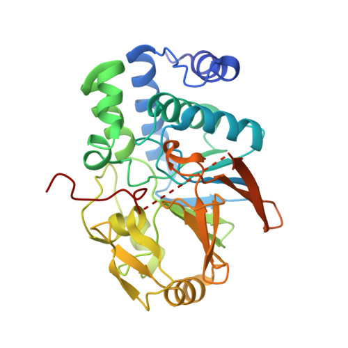
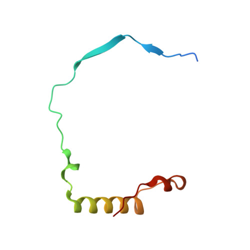
Molecular basis for substrate specificity of the Phactr1/PP1 phosphatase holoenzyme.

Fedoryshchak, R.O.Prechova, M.Butler, A.Lee, R.O'Reilly, N.Flynn, H.R.Snijders, A.P.Eder, N.Ultanir, S.Mouilleron, S.Treisman, R.

(2020) Elife 9

  • DOI: https://doi.org/10.7554/eLife.61509
  • Primary Citation Related Structures: 
    6ZEE, 6ZEF, 6ZEG, 6ZEH, 6ZEI, 6ZEJ

  • PubMed Abstract: 

    PPP-family phosphatases such as PP1 have little intrinsic specificity. Cofactors can target PP1 to substrates or subcellular locations, but it remains unclear how they might confer sequence-specificity on PP1. The cytoskeletal regulator Phactr1 is a neuronally enriched PP1 cofactor that is controlled by G-actin. Structural analysis showed that Phactr1 binding remodels PP1's hydrophobic groove, creating a new composite surface adjacent to the catalytic site. Using phosphoproteomics, we identified mouse fibroblast and neuronal Phactr1/PP1 substrates, which include cytoskeletal components and regulators. We determined high-resolution structures of Phactr1/PP1 bound to the dephosphorylated forms of its substrates IRSp53 and spectrin αII. Inversion of the phosphate in these holoenzyme-product complexes supports the proposed PPP-family catalytic mechanism. Substrate sequences C-terminal to the dephosphorylation site make intimate contacts with the composite Phactr1/PP1 surface, which are required for efficient dephosphorylation. Sequence specificity explains why Phactr1/PP1 exhibits orders-of-magnitude enhanced reactivity towards its substrates, compared to apo-PP1 or other PP1 holoenzymes.


  • Organizational Affiliation
    • Signalling and Transcription Laboratory, The Francis Crick Institute, London, United Kingdom.

Macromolecule Content 

  • Total Structure Weight: 92.14 kDa 
  • Atom Count: 6,778 
  • Modeled Residue Count: 742 
  • Deposited Residue Count: 812 
  • Unique protein chains: 2

Macromolecules

Find similar proteins by:|  3D Structure
Entity ID: 1
MoleculeChains  Sequence LengthOrganismDetailsImage
Serine/threonine-protein phosphatase PP1-alpha catalytic subunit,Spectrin alpha chain, non-erythrocytic 1A,
C [auth B]
336Homo sapiensMutation(s): 0 
Gene Names: PPP1CAPPP1ASPTAN1NEASSPTA2
EC: 3.1.3.16
UniProt & NIH Common Fund Data Resources
Find proteins for P62136 (Homo sapiens)
Explore P62136 
Go to UniProtKB:  P62136
PHAROS:  P62136
GTEx:  ENSG00000172531 
Find proteins for Q13813 (Homo sapiens)
Explore Q13813 
Go to UniProtKB:  Q13813
PHAROS:  Q13813
GTEx:  ENSG00000197694 
Entity Groups
Sequence Clusters30% Identity50% Identity70% Identity90% Identity95% Identity100% Identity
UniProt GroupsQ13813P62136
Sequence Annotations
Expand
Reference Sequence
Find similar proteins by:|  3D Structure
Entity ID: 2
MoleculeChains  Sequence LengthOrganismDetailsImage
Phosphatase and actin regulatorB [auth C],
D
70Homo sapiensMutation(s): 0 
Gene Names: PHACTR1hCG_1818446
UniProt & NIH Common Fund Data Resources
Find proteins for Q9C0D0 (Homo sapiens)
Explore Q9C0D0 
Go to UniProtKB:  Q9C0D0
PHAROS:  Q9C0D0
GTEx:  ENSG00000112137 
Entity Groups
Sequence Clusters30% Identity50% Identity70% Identity90% Identity95% Identity100% Identity
UniProt GroupQ9C0D0
Sequence Annotations
Expand
Reference Sequence

Small Molecules

Ligands 3 Unique
IDChains Name / Formula / InChI Key2D Diagram3D Interactions
SO4

Query on SO4


Download:Ideal Coordinates CCD File
K [auth B]SULFATE ION
O4 S
QAOWNCQODCNURD-UHFFFAOYSA-L
PO4

Query on PO4


Download:Ideal Coordinates CCD File
G [auth A],
J [auth B]
PHOSPHATE ION
O4 P
NBIIXXVUZAFLBC-UHFFFAOYSA-K
MN

Query on MN


Download:Ideal Coordinates CCD File
E [auth A],
F [auth A],
H [auth B],
I [auth B]
MANGANESE (II) ION
Mn
WAEMQWOKJMHJLA-UHFFFAOYSA-N

Experimental Data & Validation

Experimental Data

  • Method: X-RAY DIFFRACTION
  • Resolution: 1.30 Å
  • R-Value Free:  0.169 (Depositor), 0.169 (DCC) 
  • R-Value Work:  0.137 (Depositor), 0.137 (DCC) 
  • R-Value Observed: 0.138 (Depositor) 
Space Group: P 1 21 1
Unit Cell:
Length ( Å )Angle ( ˚ )
a = 48.501α = 90
b = 122.388β = 92.179
c = 69.343γ = 90
Software Package:
Software NamePurpose
PHENIXrefinement
PHENIXrefinement
PDB_EXTRACTdata extraction
xia2data reduction
Aimlessdata scaling
PHASERphasing

Structure Validation

View Full Validation Report


Entry History 

Deposition Data

Revision History  (Full details and data files)

  • Version 1.0: 2020-09-30
    Type: Initial release
  • Version 1.1: 2020-10-07
    Changes: Database references
  • Version 1.2: 2024-01-24
    Changes: Data collection, Database references, Refinement description