6ZDH | pdb_00006zdh

SARS-CoV-2 Spike glycoprotein in complex with a neutralizing antibody EY6A Fab


Experimental Data Snapshot

  • Method: ELECTRON MICROSCOPY
  • Resolution: 3.70 Å
  • Aggregation State: PARTICLE 
  • Reconstruction Method: SINGLE PARTICLE 

wwPDB Validation 3D Report Full Report

Validation slider image for 6ZDH

This is version 2.6 of the entry. See complete history

Literature

Structural basis for the neutralization of SARS-CoV-2 by an antibody from a convalescent patient.

Zhou, D.Duyvesteyn, H.M.E.Chen, C.P.Huang, C.G.Chen, T.H.Shih, S.R.Lin, Y.C.Cheng, C.Y.Cheng, S.H.Huang, Y.C.Lin, T.Y.Ma, C.Huo, J.Carrique, L.Malinauskas, T.Ruza, R.R.Shah, P.N.M.Tan, T.K.Rijal, P.Donat, R.F.Godwin, K.Buttigieg, K.R.Tree, J.A.Radecke, J.Paterson, N.G.Supasa, P.Mongkolsapaya, J.Screaton, G.R.Carroll, M.W.Gilbert-Jaramillo, J.Knight, M.L.James, W.Owens, R.J.Naismith, J.H.Townsend, A.R.Fry, E.E.Zhao, Y.Ren, J.Stuart, D.I.Huang, K.A.

(2020) Nat Struct Mol Biol 27: 950-958

  • DOI: https://doi.org/10.1038/s41594-020-0480-y
  • Primary Citation Related Structures: 
    6ZCZ, 6ZDG, 6ZDH, 6ZER, 6ZFO

  • PubMed Abstract: 

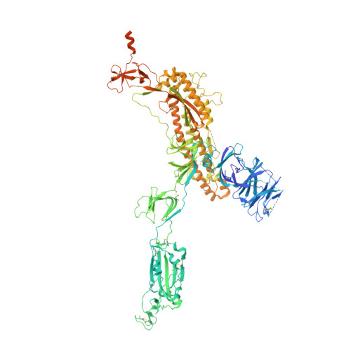
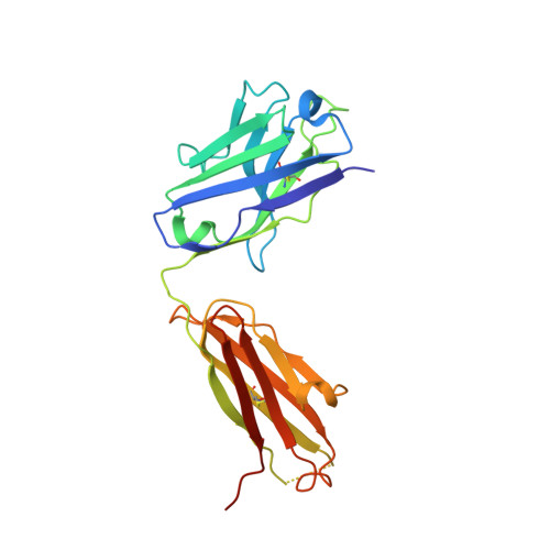
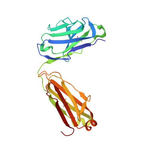
    The COVID-19 pandemic has had an unprecedented health and economic impact and there are currently no approved therapies. We have isolated an antibody, EY6A, from an individual convalescing from COVID-19 and have shown that it neutralizes SARS-CoV-2 and cross-reacts with SARS-CoV-1. EY6A Fab binds the receptor binding domain (RBD) of the viral spike glycoprotein tightly (K D of 2 nM), and a 2.6-Å-resolution crystal structure of an RBD-EY6A Fab complex identifies the highly conserved epitope, away from the ACE2 receptor binding site. Residues within this footprint are key to stabilizing the pre-fusion spike. Cryo-EM analyses of the pre-fusion spike incubated with EY6A Fab reveal a complex of the intact spike trimer with three Fabs bound and two further multimeric forms comprising the destabilized spike attached to Fab. EY6A binds what is probably a major neutralizing epitope, making it a candidate therapeutic for COVID-19.


  • Organizational Affiliation
    • Division of Structural Biology, Nuffield Department of Medicine, University of Oxford, The Wellcome Centre for Human Genetics, Headington, Oxford, UK.

Macromolecule Content 

  • Total Structure Weight: 580.79 kDa 
  • Atom Count: 34,058 
  • Modeled Residue Count: 4,308 
  • Deposited Residue Count: 5,187 
  • Unique protein chains: 3

Macromolecules

Find similar proteins by:|  3D Structure
Entity ID: 1
MoleculeChains  Sequence LengthOrganismDetailsImage
Spike glycoprotein
A, B, C
1,288Severe acute respiratory syndrome coronavirus 2Mutation(s): 5 
Gene Names: S2
UniProt
Find proteins for P0DTC2 (Severe acute respiratory syndrome coronavirus 2)
Explore P0DTC2 
Go to UniProtKB:  P0DTC2
Entity Groups
Sequence Clusters30% Identity50% Identity70% Identity90% Identity95% Identity100% Identity
UniProt GroupP0DTC2
Glycosylation
Glycosylation Sites: 12Go to GlyGen: P0DTC2-1
Sequence Annotations
Expand
Reference Sequence
Find similar proteins by:|  3D Structure
Entity ID: 2
MoleculeChains  Sequence LengthOrganismDetailsImage
EY6A heavy chainD [auth H],
F [auth D],
H [auth G]
226Homo sapiensMutation(s): 0 
Find similar proteins by:|  3D Structure
Entity ID: 3
MoleculeChains  Sequence LengthOrganismDetailsImage
EY6A light chainE [auth L],
G [auth F],
I
215Homo sapiensMutation(s): 0 

Oligosaccharides

Help  
Entity ID: 4
MoleculeChains Length2D Diagram GlycosylationD Interactions
2-acetamido-2-deoxy-beta-D-glucopyranose-(1-4)-2-acetamido-2-deoxy-beta-D-glucopyranose
J [auth E],
K [auth J],
L [auth K],
M,
N,
J [auth E],
K [auth J],
L [auth K],
M,
N,
O,
P,
Q,
R,
S,
T,
U,
V
2N-Glycosylation
Glycosylation Resources
GlyTouCan: G42666HT
GlyCosmos: G42666HT
GlyGen: G42666HT

Small Molecules

Ligands 1 Unique
IDChains Name / Formula / InChI Key2D Diagram3D Interactions
NAG

Query on NAG


Download:Ideal Coordinates CCD File
AA [auth A]
BA [auth A]
CA [auth A]
DA [auth A]
EA [auth B]
AA [auth A],
BA [auth A],
CA [auth A],
DA [auth A],
EA [auth B],
FA [auth B],
GA [auth B],
HA [auth B],
IA [auth B],
JA [auth B],
KA [auth B],
LA [auth B],
MA [auth C],
NA [auth C],
OA [auth C],
PA [auth C],
QA [auth C],
RA [auth C],
SA [auth C],
W [auth A],
X [auth A],
Y [auth A],
Z [auth A]
2-acetamido-2-deoxy-beta-D-glucopyranose
C8 H15 N O6
OVRNDRQMDRJTHS-FMDGEEDCSA-N

Experimental Data & Validation

Experimental Data

  • Method: ELECTRON MICROSCOPY
  • Resolution: 3.70 Å
  • Aggregation State: PARTICLE 
  • Reconstruction Method: SINGLE PARTICLE 
EM Software:
TaskSoftware PackageVersion
MODEL REFINEMENTPHENIX
RECONSTRUCTIONcryoSPARC

Structure Validation

View Full Validation Report


Entry History 

& Funding Information

Deposition Data


Funding OrganizationLocationGrant Number
Medical Research Council (MRC, United Kingdom)United KingdomMR/N00065X/1
Wellcome TrustUnited Kingdom101122/Z/13/Z
CAMS Innovation Fund for Medical Sciences (CIFMS)China2018-I2M-2-002

Revision History  (Full details and data files)

  • Version 1.0: 2020-07-01
    Type: Initial release
  • Version 2.0: 2020-07-29
    Type: Remediation
    Reason: Carbohydrate remediation
    Changes: Advisory, Atomic model, Data collection, Derived calculations, Structure summary
  • Version 2.1: 2020-08-12
    Changes: Database references, Structure summary
  • Version 2.2: 2020-10-21
    Changes: Database references
  • Version 2.3: 2021-02-10
    Changes: Database references
  • Version 2.4: 2021-12-22
    Changes: Database references
  • Version 2.5: 2024-10-23
    Changes: Data collection, Structure summary
  • Version 2.6: 2025-07-02
    Changes: Data collection