6Z7T | pdb_00006z7t

Nucleotide-free Myosin-II motor domain


Experimental Data Snapshot

  • Method: X-RAY DIFFRACTION
  • Resolution: 1.88 Å
  • R-Value Free: 
    0.217 (Depositor), 0.222 (DCC) 
  • R-Value Work: 
    0.191 (Depositor), 0.193 (DCC) 
  • R-Value Observed: 
    0.191 (Depositor) 

Starting Model: experimental
View more details

wwPDB Validation 3D Report Full Report

Validation slider image for 6Z7T

This is version 1.2 of the entry. See complete history

Literature

Structural and Computational Insights into a Blebbistatin-Bound Myosin•ADP Complex with Characteristics of an ADP-Release Conformation along the Two-Step Myosin Power Stoke.

Ewert, W.Franz, P.Tsiavaliaris, G.Preller, M.

(2020) Int J Mol Sci 21

  • DOI: https://doi.org/10.3390/ijms21197417
  • Primary Citation Related Structures: 
    6Z7T, 6Z7U

  • PubMed Abstract: 

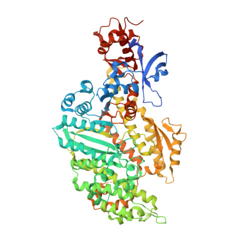
    The motor protein myosin drives a wide range of cellular and muscular functions by generating directed movement and force, fueled through adenosine triphosphate (ATP) hydrolysis. Release of the hydrolysis product adenosine diphosphate (ADP) is a fundamental and regulatory process during force production. However, details about the molecular mechanism accompanying ADP release are scarce due to the lack of representative structures. Here we solved a novel blebbistatin-bound myosin conformation with critical structural elements in positions between the myosin pre-power stroke and rigor states. ADP in this structure is repositioned towards the surface by the phosphate-sensing P-loop, and stabilized in a partially unbound conformation via a salt-bridge between Arg131 and Glu187. A 5 Å rotation separates the mechanical converter in this conformation from the rigor position. The crystallized myosin structure thus resembles a conformation towards the end of the two-step power stroke, associated with ADP release. Computationally reconstructing ADP release from myosin by means of molecular dynamics simulations further supported the existence of an equivalent conformation along the power stroke that shows the same major characteristics in the myosin motor domain as the resolved blebbistatin-bound myosin-II·ADP crystal structure, and identified a communication hub centered on Arg232 that mediates chemomechanical energy transduction.


  • Organizational Affiliation
    • Institute for Biophysical Chemistry, Structural Bioinformatics and Chemical Biology, Hannover Medical School, 30625 Hannover, Germany.

Macromolecule Content 

  • Total Structure Weight: 182.54 kDa 
  • Atom Count: 12,958 
  • Modeled Residue Count: 1,456 
  • Deposited Residue Count: 1,576 
  • Unique protein chains: 1

Macromolecules

Find similar proteins by:|  3D Structure
Entity ID: 1
MoleculeChains  Sequence LengthOrganismDetailsImage
Myosin-2 heavy chain
A, B
788Dictyostelium discoideumMutation(s): 0 
Gene Names: mhcADDB_G0286355
UniProt
Find proteins for P08799 (Dictyostelium discoideum)
Explore P08799 
Go to UniProtKB:  P08799
Entity Groups
Sequence Clusters30% Identity50% Identity70% Identity90% Identity95% Identity100% Identity
UniProt GroupP08799
Sequence Annotations
Expand
Reference Sequence

Small Molecules

Ligands 5 Unique
IDChains Name / Formula / InChI Key2D Diagram3D Interactions
PGE

Query on PGE


Download:Ideal Coordinates CCD File
KA [auth B]TRIETHYLENE GLYCOL
C6 H14 O4
ZIBGPFATKBEMQZ-UHFFFAOYSA-N
PEG

Query on PEG


Download:Ideal Coordinates CCD File
LA [auth B]DI(HYDROXYETHYL)ETHER
C4 H10 O3
MTHSVFCYNBDYFN-UHFFFAOYSA-N
GOL

Query on GOL


Download:Ideal Coordinates CCD File
FA [auth B]
GA [auth B]
HA [auth B]
IA [auth B]
JA [auth B]
FA [auth B],
GA [auth B],
HA [auth B],
IA [auth B],
JA [auth B],
O [auth A],
P [auth A],
Q [auth A],
R [auth A],
S [auth A]
GLYCEROL
C3 H8 O3
PEDCQBHIVMGVHV-UHFFFAOYSA-N
EDO

Query on EDO


Download:Ideal Coordinates CCD File
AA [auth B]
BA [auth B]
CA [auth B]
DA [auth B]
E [auth A]
AA [auth B],
BA [auth B],
CA [auth B],
DA [auth B],
E [auth A],
EA [auth B],
F [auth A],
G [auth A],
H [auth A],
I [auth A],
J [auth A],
K [auth A],
L [auth A],
M [auth A],
N [auth A],
V [auth B],
W [auth B],
X [auth B],
Y [auth B],
Z [auth B]
1,2-ETHANEDIOL
C2 H6 O2
LYCAIKOWRPUZTN-UHFFFAOYSA-N
MG

Query on MG


Download:Ideal Coordinates CCD File
C [auth A],
D [auth A],
T [auth B],
U [auth B]
MAGNESIUM ION
Mg
JLVVSXFLKOJNIY-UHFFFAOYSA-N

Experimental Data & Validation

Experimental Data

  • Method: X-RAY DIFFRACTION
  • Resolution: 1.88 Å
  • R-Value Free:  0.217 (Depositor), 0.222 (DCC) 
  • R-Value Work:  0.191 (Depositor), 0.193 (DCC) 
  • R-Value Observed: 0.191 (Depositor) 
Space Group: P 1 21 1
Unit Cell:
Length ( Å )Angle ( ˚ )
a = 56.681α = 90
b = 174.395β = 106.384
c = 100.252γ = 90
Software Package:
Software NamePurpose
PHENIXrefinement
XDSdata reduction
Aimlessdata scaling
PHASERphasing
Cootmodel building

Structure Validation

View Full Validation Report


Entry History 

& Funding Information

Deposition Data


Funding OrganizationLocationGrant Number
German Research Foundation (DFG)GermanyPR 1478/2-1

Revision History  (Full details and data files)

  • Version 1.0: 2020-10-21
    Type: Initial release
  • Version 1.1: 2020-10-28
    Changes: Database references
  • Version 1.2: 2024-01-24
    Changes: Data collection, Database references, Refinement description