


Crystal structure of Aurora A (STK6) in complex with macrocycle ODS2003208
Chaikuad, A., Benderitter, P., Hoflack, J., Denis, A., Knapp, S., Structural Genomics Consortium (SGC)To be published.
Experimental Data Snapshot
Starting Model: experimental
View more details
Entity ID: 1 | |||||
|---|---|---|---|---|---|
| Molecule | Chains | Sequence Length | Organism | Details | Image |
| Aurora kinase A | 285 | Homo sapiens | Mutation(s): 0 Gene Names: AURKA, AIK, AIRK1, ARK1, AURA, AYK1, BTAK, IAK1, STK15, STK6 EC: 2.7.11.1 | 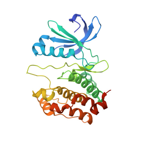 | |
UniProt & NIH Common Fund Data Resources | |||||
PHAROS: O14965 GTEx: ENSG00000087586 | |||||
Entity Groups | |||||
| Sequence Clusters | 30% Identity50% Identity70% Identity90% Identity95% Identity100% Identity | ||||
| UniProt Group | O14965 | ||||
Sequence AnnotationsExpand | |||||
Reference Sequence | |||||
| Ligands 3 Unique | |||||
|---|---|---|---|---|---|
| ID | Chains | Name / Formula / InChI Key | 2D Diagram | 3D Interactions | |
| Q7Q (Subject of Investigation/LOI) Download:Ideal Coordinates CCD File | I [auth A] | 6-(2-methoxyethoxy)-11-methyl-8-oxa-2,11,15,19,21,23-hexazatetracyclo[15.6.1.13,7.020,24]pentacosa-1(23),3(25),4,6,17,20(24),21-heptaen-10-one C22 H28 N6 O4 MPVLMOBPNHBSRH-UHFFFAOYSA-N |  | ||
| CIT Download:Ideal Coordinates CCD File | B [auth A], C [auth A] | CITRIC ACID C6 H8 O7 KRKNYBCHXYNGOX-UHFFFAOYSA-N |  | ||
| EDO Download:Ideal Coordinates CCD File | D [auth A], E [auth A], F [auth A], G [auth A], H [auth A] | 1,2-ETHANEDIOL C2 H6 O2 LYCAIKOWRPUZTN-UHFFFAOYSA-N |  | ||
| Length ( Å ) | Angle ( ˚ ) |
|---|---|
| a = 82.83 | α = 90 |
| b = 82.83 | β = 90 |
| c = 173.301 | γ = 120 |
| Software Name | Purpose |
|---|---|
| SCALA | data scaling |
| REFMAC | refinement |
| PDB_EXTRACT | data extraction |
| iMOSFLM | data reduction |
| PHASER | phasing |


