6Z4T | pdb_00006z4t

sperm whale myoglobin mutant (H64V V64A) bearing the non-canonical amino acid 2-Amino-3-(thiazol-5-yl)propanoic acid as axial heme ligand


Experimental Data Snapshot

  • Method: X-RAY DIFFRACTION
  • Resolution: 1.23 Å
  • R-Value Free: 
    0.188 (Depositor), 0.187 (DCC) 
  • R-Value Work: 
    0.150 (Depositor), 0.151 (DCC) 
  • R-Value Observed: 
    0.152 (Depositor) 

Starting Model: experimental
View more details

wwPDB Validation 3D Report Full Report

Validation slider image for 6Z4T

Ligand Structure Quality Assessment 


This is version 3.0 of the entry. See complete history

Literature

Noncanonical Heme Ligands Steer Carbene Transfer Reactivity in an Artificial Metalloenzyme*.

Pott, M.Tinzl, M.Hayashi, T.Ota, Y.Dunkelmann, D.Mittl, P.R.E.Hilvert, D.

(2021) Angew Chem Int Ed Engl 60: 15063-15068

  • DOI: https://doi.org/10.1002/anie.202103437
  • Primary Citation Related Structures: 
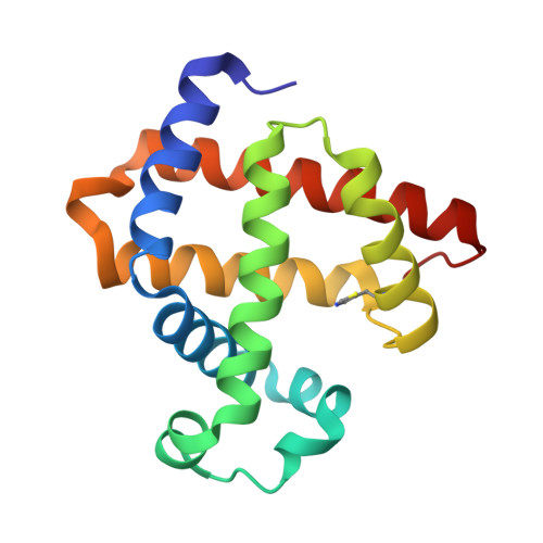
    6Z4R, 6Z4T

  • PubMed Abstract: 

    Changing the primary metal coordination sphere is a powerful strategy for tuning metalloprotein properties. Here we used amber stop codon suppression with engineered pyrrolysyl-tRNA synthetases, including two newly evolved enzymes, to replace the proximal histidine in myoglobin with N δ -methylhistidine, 5-thiazoylalanine, 4-thiazoylalanine and 3-(3-thienyl)alanine. In addition to tuning the heme redox potential over a >200 mV range, these noncanonical ligands modulate the protein's carbene transfer activity with ethyl diazoacetate. Variants with increased reduction potential proved superior for cyclopropanation and N-H insertion, whereas variants with reduced E o values gave higher S-H insertion activity. Given the functional importance of histidine in many enzymes, these genetically encoded analogues could be valuable tools for probing mechanism and enabling new chemistries.


  • Organizational Affiliation
    • Laboratory of Organic Chemistry, ETH Zürich, 8093, Zürich, Switzerland.

Macromolecule Content 

  • Total Structure Weight: 17.89 kDa 
  • Atom Count: 1,518 
  • Modeled Residue Count: 154 
  • Deposited Residue Count: 154 
  • Unique protein chains: 1

Macromolecules

Find similar proteins by:|  3D Structure
Entity ID: 1
MoleculeChains  Sequence LengthOrganismDetailsImage
Myoglobin154Physeter macrocephalusMutation(s): 3 
Gene Names: MB
EC: 1.11.1 (UniProt), 1.7 (UniProt)
UniProt
Find proteins for P02185 (Physeter macrocephalus)
Explore P02185 
Go to UniProtKB:  P02185
Entity Groups
Sequence Clusters30% Identity50% Identity70% Identity90% Identity95% Identity100% Identity
UniProt GroupP02185
Sequence Annotations
Expand
Reference Sequence

Small Molecules

Modified Residues  1 Unique
IDChains TypeFormula2D DiagramParent
Q75
Query on Q75
A
L-PEPTIDE LINKINGC6 H8 N2 O2 SHIS

Experimental Data & Validation

Experimental Data

  • Method: X-RAY DIFFRACTION
  • Resolution: 1.23 Å
  • R-Value Free:  0.188 (Depositor), 0.187 (DCC) 
  • R-Value Work:  0.150 (Depositor), 0.151 (DCC) 
  • R-Value Observed: 0.152 (Depositor) 
Space Group: P 21 21 21
Unit Cell:
Length ( Å )Angle ( ˚ )
a = 40.009α = 90
b = 47.327β = 90
c = 76.897γ = 90
Software Package:
Software NamePurpose
XDSdata reduction
Aimlessdata scaling
PHENIXmodel building
PHENIXphasing
PHENIXrefinement

Structure Validation

View Full Validation Report


Ligand Structure Quality Assessment 


Entry History 

Deposition Data

Revision History  (Full details and data files)

  • Version 1.0: 2021-04-28
    Type: Initial release
  • Version 1.1: 2021-06-30
    Changes: Database references
  • Version 2.0: 2023-09-13
    Changes: Atomic model, Data collection, Database references, Derived calculations, Non-polymer description, Polymer sequence, Structure summary
  • Version 2.1: 2024-01-24
    Changes: Refinement description
  • Version 3.0: 2025-07-30
    Changes: Polymer sequence, Structure summary