6Z4S | pdb_00006z4s

Crystal structure of the neurotensin receptor 1 (NTSR1-H4bmx) in complex with the small molecule inverse agonist SR48692


Experimental Data Snapshot

  • Method: X-RAY DIFFRACTION
  • Resolution: 2.71 Å
  • R-Value Free: 
    0.289 (Depositor), 0.295 (DCC) 
  • R-Value Work: 
    0.273 (Depositor), 0.275 (DCC) 

Starting Model: experimental
View more details

wwPDB Validation 3D Report Full Report

Validation slider image for 6Z4S

Ligand Structure Quality Assessment 


This is version 1.4 of the entry. See complete history

Literature

Complexes of the neurotensin receptor 1 with small-molecule ligands reveal structural determinants of full, partial, and inverse agonism.

Deluigi, M.Klipp, A.Klenk, C.Merklinger, L.Eberle, S.A.Morstein, L.Heine, P.Mittl, P.R.E.Ernst, P.Kamenecka, T.M.He, Y.Vacca, S.Egloff, P.Honegger, A.Pluckthun, A.

(2021) Sci Adv 7

  • DOI: https://doi.org/10.1126/sciadv.abe5504
  • Primary Citation Related Structures: 
    6YVR, 6Z4Q, 6Z4S, 6Z4V, 6Z66, 6Z8N, 6ZA8, 6ZIN

  • PubMed Abstract: 

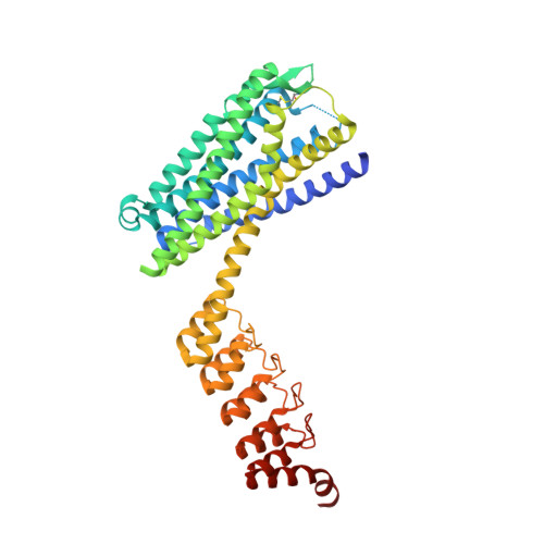
    Neurotensin receptor 1 (NTSR1) and related G protein-coupled receptors of the ghrelin family are clinically unexploited, and several mechanistic aspects of their activation and inactivation have remained unclear. Enabled by a new crystallization design, we present five new structures: apo-state NTSR1 as well as complexes with nonpeptide inverse agonists SR48692 and SR142948A, partial agonist RTI-3a, and the novel full agonist SRI-9829, providing structural rationales on how ligands modulate NTSR1. The inverse agonists favor a large extracellular opening of helices VI and VII, undescribed so far for NTSR1, causing a constriction of the intracellular portion. In contrast, the full and partial agonists induce a binding site contraction, and their efficacy correlates with the ability to mimic the binding mode of the endogenous agonist neurotensin. Providing evidence of helical and side-chain rearrangements modulating receptor activation, our structural and functional data expand the mechanistic understanding of NTSR1 and potentially other peptidergic receptors.


  • Organizational Affiliation
    • Department of Biochemistry, University of Zurich, Winterthurerstrasse 190, CH-8057 Zurich, Switzerland.

Macromolecule Content 

  • Total Structure Weight: 53.74 kDa 
  • Atom Count: 3,330 
  • Modeled Residue Count: 448 
  • Deposited Residue Count: 482 
  • Unique protein chains: 1

Macromolecules

Find similar proteins by:|  3D Structure
Entity ID: 1
MoleculeChains  Sequence LengthOrganismDetailsImage
Neurotensin receptor type 1,DARPin,HRV 3C protease recognition sequenceA [auth AAA]482Rattus norvegicussynthetic constructMutation(s): 22 
Gene Names: Ntsr1Ntsr
Membrane Entity: Yes 
UniProt
Find proteins for P20789 (Rattus norvegicus)
Explore P20789 
Go to UniProtKB:  P20789
Entity Groups
Sequence Clusters30% Identity50% Identity70% Identity90% Identity95% Identity100% Identity
UniProt GroupP20789
Sequence Annotations
Expand
Reference Sequence

Small Molecules

Ligands 1 Unique
IDChains Name / Formula / InChI Key2D Diagram3D Interactions
Q6Q
(Subject of Investigation/LOI)

Query on Q6Q


Download:Ideal Coordinates CCD File
B [auth AAA]2-[[1-(7-chloranylquinolin-4-yl)-5-(2,6-dimethoxyphenyl)pyrazol-3-yl]carbonylamino]adamantane-2-carboxylic acid
C32 H31 Cl N4 O5
DYLJVOXRWLXDIG-LWXMPVTCSA-N

Experimental Data & Validation

Experimental Data

  • Method: X-RAY DIFFRACTION
  • Resolution: 2.71 Å
  • R-Value Free:  0.289 (Depositor), 0.295 (DCC) 
  • R-Value Work:  0.273 (Depositor), 0.275 (DCC) 
Space Group: P 21 21 21
Unit Cell:
Length ( Å )Angle ( ˚ )
a = 43.518α = 90
b = 77.727β = 90
c = 158.394γ = 90
Software Package:
Software NamePurpose
REFMACrefinement
XDSdata reduction
Aimlessdata scaling
PHASERphasing

Structure Validation

View Full Validation Report


Ligand Structure Quality Assessment 


Entry History 

& Funding Information

Deposition Data


Funding OrganizationLocationGrant Number
Swiss National Science FoundationSwitzerland31003A_182334

Revision History  (Full details and data files)

  • Version 1.0: 2021-02-10
    Type: Initial release
  • Version 1.1: 2021-02-24
    Changes: Database references
  • Version 1.2: 2021-03-17
    Changes: Database references
  • Version 1.3: 2024-01-24
    Changes: Data collection, Database references, Derived calculations, Refinement description
  • Version 1.4: 2024-11-06
    Changes: Structure summary