6Z3B | pdb_00006z3b

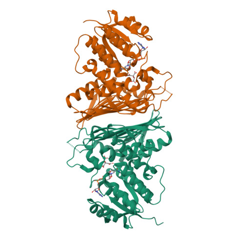
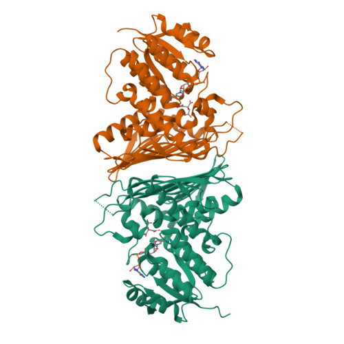
Low resolution structure of RgNanOx


Experimental Data Snapshot

  • Method: X-RAY DIFFRACTION
  • Resolution: 2.58 Å
  • R-Value Free: 
    0.250 (Depositor), 0.260 (DCC) 
  • R-Value Work: 
    0.212 (Depositor), 0.220 (DCC) 
  • R-Value Observed: 
    0.214 (Depositor) 

Starting Model: experimental
View more details

wwPDB Validation   3D Report Full Report


Ligand Structure Quality Assessment 

Created with Raphaël 2.3.0Worse 01 BetterLigand structure goodness of fit to experimental dataBest fitted NADClick on this verticalbar to view details

This is version 1.2 of the entry. See complete history


Literature

Uncovering a novel molecular mechanism for scavenging sialic acids in bacteria.

Bell, A.Severi, E.Lee, M.Monaco, S.Latousakis, D.Angulo, J.Thomas, G.H.Naismith, J.H.Juge, N.

(2020) J Biological Chem 295: 13724-13736

  • DOI: https://doi.org/10.1074/jbc.RA120.014454
  • Primary Citation of Related Structures:  
    6Z3B, 6Z3C

  • PubMed Abstract: 

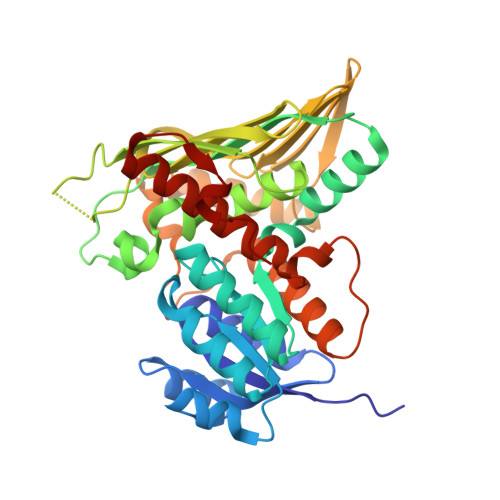
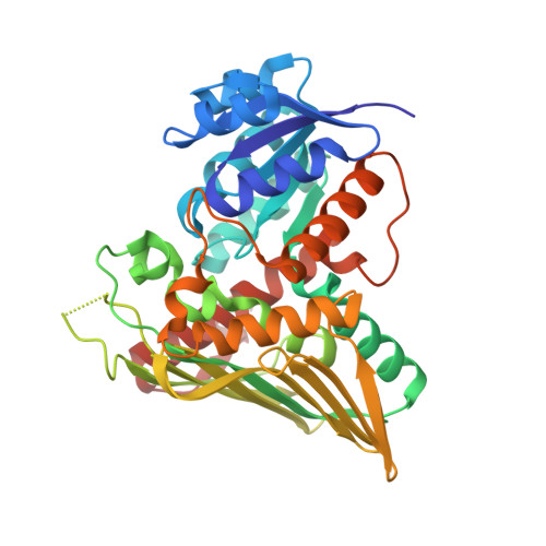
    The human gut symbiont Ruminococcus gnavus scavenges host-derived N -acetylneuraminic acid (Neu5Ac) from mucins by converting it to 2,7-anhydro-Neu5Ac. We previously showed that 2,7-anhydro-Neu5Ac is transported into R. gnavus ATCC 29149 before being converted back to Neu5Ac for further metabolic processing. However, the molecular mechanism leading to the conversion of 2,7-anhydro-Neu5Ac to Neu5Ac remained elusive. Using 1D and 2D NMR, we elucidated the multistep enzymatic mechanism of the oxidoreductase ( Rg NanOx) that leads to the reversible conversion of 2,7-anhydro-Neu5Ac to Neu5Ac through formation of a 4-keto-2-deoxy-2,3-dehydro- N -acetylneuraminic acid intermediate and NAD + regeneration. The crystal structure of Rg NanOx in complex with the NAD + cofactor showed a protein dimer with a Rossman fold. Guided by the Rg NanOx structure, we identified catalytic residues by site-directed mutagenesis. Bioinformatics analyses revealed the presence of Rg NanOx homologues across Gram-negative and Gram-positive bacterial species and co-occurrence with sialic acid transporters. We showed by electrospray ionization spray MS that the Escherichia coli homologue YjhC displayed activity against 2,7-anhydro-Neu5Ac and that E. coli could catabolize 2,7-anhydro-Neu5Ac. Differential scanning fluorimetry analyses confirmed the binding of YjhC to the substrates 2,7-anhydro-Neu5Ac and Neu5Ac, as well as to co-factors NAD and NADH. Finally, using E. coli mutants and complementation growth assays, we demonstrated that 2,7-anhydro-Neu5Ac catabolism in E. coli depended on YjhC and on the predicted sialic acid transporter YjhB. These results revealed the molecular mechanisms of 2,7-anhydro-Neu5Ac catabolism across bacterial species and a novel sialic acid transport and catabolism pathway in E. coli .


  • Organizational Affiliation

    Gut Microbes and Health Institute Strategic Programme, Quadram Institute Bioscience, Norwich, United Kingdom.


Macromolecules
Find similar proteins by:  (by identity cutoff)  |  3D Structure
Entity ID: 1
MoleculeChains Sequence LengthOrganismDetailsImage
Gfo/Idh/MocA family oxidoreductase374Mediterraneibacter gnavusMutation(s): 0 
Gene Names: CDL25_11485CDL27_13940DW270_01520DW812_00100DWY88_14550DWZ50_08505
EC: 4.2.1
UniProt
Find proteins for A7B558 (Mediterraneibacter gnavus (strain ATCC 29149 / DSM 114966 / JCM 6515 / VPI C7-9))
Explore A7B558 
Go to UniProtKB:  A7B558
Entity Groups  
Sequence Clusters30% Identity50% Identity70% Identity90% Identity95% Identity100% Identity
UniProt GroupA7B558
Sequence Annotations
Expand
  • Reference Sequence
Find similar proteins by:  (by identity cutoff)  |  3D Structure
Entity ID: 2
MoleculeChains Sequence LengthOrganismDetailsImage
Gfo/Idh/MocA family oxidoreductase369Mediterraneibacter gnavusMutation(s): 0 
Gene Names: CDL25_11485CDL27_13940DW270_01520DW812_00100DWY88_14550DWZ50_08505
EC: 4.2.1
UniProt
Find proteins for A7B558 (Mediterraneibacter gnavus (strain ATCC 29149 / DSM 114966 / JCM 6515 / VPI C7-9))
Explore A7B558 
Go to UniProtKB:  A7B558
Entity Groups  
Sequence Clusters30% Identity50% Identity70% Identity90% Identity95% Identity100% Identity
UniProt GroupA7B558
Sequence Annotations
Expand
  • Reference Sequence
Experimental Data & Validation

Experimental Data

  • Method: X-RAY DIFFRACTION
  • Resolution: 2.58 Å
  • R-Value Free:  0.250 (Depositor), 0.260 (DCC) 
  • R-Value Work:  0.212 (Depositor), 0.220 (DCC) 
  • R-Value Observed: 0.214 (Depositor) 
Space Group: I 41 2 2
Unit Cell:
Length ( Å )Angle ( ˚ )
a = 150.1α = 90
b = 150.1β = 90
c = 193.31γ = 90
Software Package:
Software NamePurpose
REFMACrefinement
Aimlessdata reduction
XSCALEdata scaling
PHASERphasing

Structure Validation

View Full Validation Report


Ligand Structure Quality Assessment 

Created with Raphaël 2.3.0Worse 01 BetterLigand structure goodness of fit to experimental dataBest fitted NADClick on this verticalbar to view details

Entry History 

Deposition Data

Revision History  (Full details and data files)

  • Version 1.0: 2020-07-22
    Type: Initial release
  • Version 1.1: 2021-02-03
    Changes: Database references
  • Version 1.2: 2024-01-24
    Changes: Data collection, Database references, Refinement description