6Z2V | pdb_00006z2v

CLK3 A319V mutant bound with beta-carboline KH-CARB13 (Cpd 3)


Experimental Data Snapshot

  • Method: X-RAY DIFFRACTION
  • Resolution: 2.60 Å
  • R-Value Free: 
    0.265 (Depositor), 0.264 (DCC) 
  • R-Value Work: 
    0.202 (Depositor), 0.209 (DCC) 
  • R-Value Observed: 
    0.205 (Depositor) 

Starting Model: experimental
View more details

wwPDB Validation 3D Report Full Report

Validation slider image for 6Z2V

Ligand Structure Quality Assessment 


This is version 1.2 of the entry. See complete history

Literature

DFG-1 Residue Controls Inhibitor Binding Mode and Affinity, Providing a Basis for Rational Design of Kinase Inhibitor Selectivity.

Schroder, M.Bullock, A.N.Fedorov, O.Bracher, F.Chaikuad, A.Knapp, S.

(2020) J Med Chem 63: 10224-10234

  • DOI: https://doi.org/10.1021/acs.jmedchem.0c00898
  • Primary Citation Related Structures: 
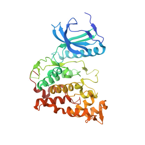
    6YTA, 6YTD, 6YTE, 6YTG, 6YTI, 6YTW, 6YTY, 6YU1, 6Z2V, 6ZLN

  • PubMed Abstract: 

    Selectivity remains a challenge for ATP-mimetic kinase inhibitors, an issue that may be overcome by targeting unique residues or binding pockets. However, to date only few strategies have been developed. Here we identify that bulky residues located N-terminal to the DFG motif (DFG-1) represent an opportunity for designing highly selective inhibitors with unexpected binding modes. We demonstrate that several diverse inhibitors exerted selective, noncanonical binding modes that exclusively target large hydrophobic DFG-1 residues present in many kinases including PIM, CK1, DAPK, and CLK. By use of the CLK family as a model, structural and biochemical data revealed that the DFG-1 valine controlled a noncanonical binding mode in CLK1, providing a rationale for selectivity over the closely related CLK3 which harbors a smaller DFG-1 alanine. Our data suggest that targeting the restricted back pocket in the small fraction of kinases that harbor bulky DFG-1 residues offers a versatile selectivity filter for inhibitor design.


  • Organizational Affiliation
    • Institute of Pharmaceutical Chemistry, Goethe-University Frankfurt, Max von Lauestraße 9, 60438 Frankfurt, Germany.

Macromolecule Content 

  • Total Structure Weight: 42.81 kDa 
  • Atom Count: 2,851 
  • Modeled Residue Count: 339 
  • Deposited Residue Count: 360 
  • Unique protein chains: 1

Macromolecules

Find similar proteins by:|  3D Structure
Entity ID: 1
MoleculeChains  Sequence LengthOrganismDetailsImage
Dual specificity protein kinase CLK3360Homo sapiensMutation(s): 1 
Gene Names: CLK3
EC: 2.7.12.1
UniProt & NIH Common Fund Data Resources
Find proteins for P49761 (Homo sapiens)
Explore P49761 
Go to UniProtKB:  P49761
PHAROS:  P49761
GTEx:  ENSG00000179335 
Entity Groups
Sequence Clusters30% Identity50% Identity70% Identity90% Identity95% Identity100% Identity
UniProt GroupP49761
Sequence Annotations
Expand
Reference Sequence

Small Molecules

Ligands 3 Unique
IDChains Name / Formula / InChI Key2D Diagram3D Interactions
KHC
(Subject of Investigation/LOI)

Query on KHC


Download:Ideal Coordinates CCD File
B [auth A](4~{S})-7,8-bis(chloranyl)-9-methyl-1-oxidanylidene-spiro[2,4-dihydropyrido[3,4-b]indole-3,4'-piperidine]-4-carbonitrile
C17 H16 Cl2 N4 O
OWAWOKKHZNRHGV-JTQLQIEISA-N
EDO

Query on EDO


Download:Ideal Coordinates CCD File
D [auth A]1,2-ETHANEDIOL
C2 H6 O2
LYCAIKOWRPUZTN-UHFFFAOYSA-N
K

Query on K


Download:Ideal Coordinates CCD File
C [auth A]POTASSIUM ION
K
NPYPAHLBTDXSSS-UHFFFAOYSA-N

Experimental Data & Validation

Experimental Data

  • Method: X-RAY DIFFRACTION
  • Resolution: 2.60 Å
  • R-Value Free:  0.265 (Depositor), 0.264 (DCC) 
  • R-Value Work:  0.202 (Depositor), 0.209 (DCC) 
  • R-Value Observed: 0.205 (Depositor) 
Space Group: I 1 2 1
Unit Cell:
Length ( Å )Angle ( ˚ )
a = 84.376α = 90
b = 45.393β = 111.16
c = 106.334γ = 90
Software Package:
Software NamePurpose
Aimlessdata scaling
REFMACrefinement
PDB_EXTRACTdata extraction
XDSdata reduction
PHASERphasing

Structure Validation

View Full Validation Report


Ligand Structure Quality Assessment 


Entry History 

Deposition Data

Revision History  (Full details and data files)

  • Version 1.0: 2020-07-29
    Type: Initial release
  • Version 1.1: 2020-10-14
    Changes: Database references
  • Version 1.2: 2024-01-24
    Changes: Data collection, Database references, Refinement description