6YX5 | pdb_00006yx5

Structure of DrrA from Legionella pneumophilia in complex with human Rab8a


Experimental Data Snapshot

  • Method: X-RAY DIFFRACTION
  • Resolution: 2.14 Å
  • R-Value Free: 
    0.238 (Depositor), 0.242 (DCC) 
  • R-Value Work: 
    0.207 (Depositor), 0.217 (DCC) 
  • R-Value Observed: 
    0.208 (Depositor) 

Starting Models: experimental
View more details

wwPDB Validation 3D Report Full Report

Validation slider image for 6YX5

Ligand Structure Quality Assessment 


This is version 1.3 of the entry. See complete history

Literature

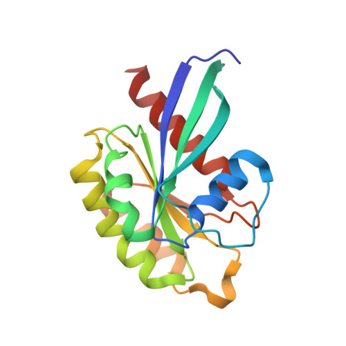
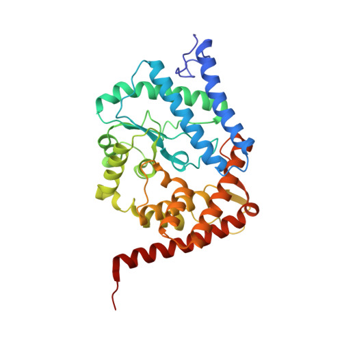
Rab1-AMPylation by Legionella DrrA is allosterically activated by Rab1.

Du, J.Wrisberg, M.V.Gulen, B.Stahl, M.Pett, C.Hedberg, C.Lang, K.Schneider, S.Itzen, A.

(2021) Nat Commun 12: 460-460

  • DOI: https://doi.org/10.1038/s41467-020-20702-2
  • Primary Citation Related Structures: 
    6YX5

  • PubMed Abstract: 

    Legionella pneumophila infects eukaryotic cells by forming a replicative organelle - the Legionella containing vacuole. During this process, the bacterial protein DrrA/SidM is secreted and manipulates the activity and post-translational modification (PTM) states of the vesicular trafficking regulator Rab1. As a result, Rab1 is modified with an adenosine monophosphate (AMP), and this process is referred to as AMPylation. Here, we use a chemical approach to stabilise low-affinity Rab:DrrA complexes in a site-specific manner to gain insight into the molecular basis of the interaction between the Rab protein and the AMPylation domain of DrrA. The crystal structure of the Rab:DrrA complex reveals a previously unknown non-conventional Rab-binding site (NC-RBS). Biochemical characterisation demonstrates allosteric stimulation of the AMPylation activity of DrrA via Rab binding to the NC-RBS. We speculate that allosteric control of DrrA could in principle prevent random and potentially cytotoxic AMPylation in the host, thereby perhaps ensuring efficient infection by Legionella.


  • Organizational Affiliation
    • Center for Integrated Protein Science Munich (CIPSM), Department of Chemistry, Technical University of Munich, Garching, 85748, Germany.

Macromolecule Content 

  • Total Structure Weight: 60.8 kDa 
  • Atom Count: 4,353 
  • Modeled Residue Count: 512 
  • Deposited Residue Count: 514 
  • Unique protein chains: 2

Macromolecules

Find similar proteins by:|  3D Structure
Entity ID: 1
MoleculeChains  Sequence LengthOrganismDetailsImage
Ras-related protein Rab-8A174Homo sapiensMutation(s): 0 
Gene Names: Rab8a
EC: 3.6.5.2
UniProt & NIH Common Fund Data Resources
Find proteins for P61006 (Homo sapiens)
Explore P61006 
Go to UniProtKB:  P61006
PHAROS:  P61006
GTEx:  ENSG00000167461 
Entity Groups
Sequence Clusters30% Identity50% Identity70% Identity90% Identity95% Identity100% Identity
UniProt GroupP61006
Sequence Annotations
Expand
Reference Sequence
Find similar proteins by:|  3D Structure
Entity ID: 2
MoleculeChains  Sequence LengthOrganismDetailsImage
Multifunctional virulence effector protein DrrA340Legionella pneumophilaMutation(s): 1 
EC: 2.7.7.108
UniProt
Find proteins for Q29ST3 (Legionella pneumophila)
Explore Q29ST3 
Go to UniProtKB:  Q29ST3
Entity Groups
Sequence Clusters30% Identity50% Identity70% Identity90% Identity95% Identity100% Identity
UniProt GroupQ29ST3
Sequence Annotations
Expand
Reference Sequence

Small Molecules

Ligands 5 Unique
IDChains Name / Formula / InChI Key2D Diagram3D Interactions
GNP

Query on GNP


Download:Ideal Coordinates CCD File
D [auth A]PHOSPHOAMINOPHOSPHONIC ACID-GUANYLATE ESTER
C10 H17 N6 O13 P3
UQABYHGXWYXDTK-UUOKFMHZSA-N
LJN

Query on LJN


Download:Ideal Coordinates CCD File
F [auth A][(2~{R},3~{S},4~{R},5~{R})-5-[4-(acetamidomethyl)-1,2,3-triazol-1-yl]-3,4-bis(oxidanyl)oxolan-2-yl]methyl dihydrogen phosphate
C10 H17 N4 O8 P
WKSDLIPBAPRDGN-ZYUZMQFOSA-N
GLC

Query on GLC


Download:Ideal Coordinates CCD File
E [auth A],
I [auth B],
J [auth B]
alpha-D-glucopyranose
C6 H12 O6
WQZGKKKJIJFFOK-DVKNGEFBSA-N
SO4

Query on SO4


Download:Ideal Coordinates CCD File
G [auth A],
H [auth A],
K [auth B],
L [auth B],
M [auth B]
SULFATE ION
O4 S
QAOWNCQODCNURD-UHFFFAOYSA-L
MG

Query on MG


Download:Ideal Coordinates CCD File
C [auth A]MAGNESIUM ION
Mg
JLVVSXFLKOJNIY-UHFFFAOYSA-N

Experimental Data & Validation

Experimental Data

  • Method: X-RAY DIFFRACTION
  • Resolution: 2.14 Å
  • R-Value Free:  0.238 (Depositor), 0.242 (DCC) 
  • R-Value Work:  0.207 (Depositor), 0.217 (DCC) 
  • R-Value Observed: 0.208 (Depositor) 
Space Group: P 3 2 1
Unit Cell:
Length ( Å )Angle ( ˚ )
a = 142.309α = 90
b = 142.309β = 90
c = 76.616γ = 120
Software Package:
Software NamePurpose
XDSdata reduction
REFMACrefinement
PDB_EXTRACTdata extraction
XDSdata scaling
PHASERphasing

Structure Validation

View Full Validation Report


Ligand Structure Quality Assessment 


Entry History 

& Funding Information

Deposition Data


Funding OrganizationLocationGrant Number
German Research Foundation (DFG)GermanySFB1035
German Research Foundation (DFG)GermanyEXC114

Revision History  (Full details and data files)

  • Version 1.0: 2020-12-23
    Type: Initial release
  • Version 1.1: 2021-02-03
    Changes: Database references
  • Version 1.2: 2024-01-24
    Changes: Data collection, Database references, Refinement description
  • Version 1.3: 2024-11-20
    Changes: Structure summary