6YQQ | pdb_00006yqq

ForT-PRPP complex


Experimental Data Snapshot

  • Method: X-RAY DIFFRACTION
  • Resolution: 2.50 Å
  • R-Value Free: 
    0.265 (Depositor), 0.291 (DCC) 
  • R-Value Work: 
    0.225 (Depositor), 0.257 (DCC) 
  • R-Value Observed: 
    0.227 (Depositor) 

Starting Model: other
View more details

wwPDB Validation 3D Report Full Report

Validation slider image for 6YQQ

Ligand Structure Quality Assessment 


This is version 1.4 of the entry. See complete history

Literature

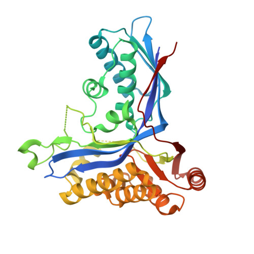
Uncovering the chemistry of C-C bond formation in C-nucleoside biosynthesis: crystal structure of a C-glycoside synthase/PRPP complex.

Gao, S.Radadiya, A.Li, W.Liu, H.Zhu, W.de Crecy-Lagard, V.Richards, N.G.J.Naismith, J.H.

(2020) Chem Commun (Camb) 56: 7617-7620

  • DOI: https://doi.org/10.1039/d0cc02834g
  • Primary Citation Related Structures: 
    6YQQ

  • PubMed Abstract: 

    The enzyme ForT catalyzes C-C bond formation between 5'-phosphoribosyl-1'-pyrophosphate (PRPP) and 4-amino-1H-pyrazole-3,5-dicarboxylate to make a key intermediate in the biosynthesis of formycin A 5'-phosphate by Streptomyces kaniharaensis. We report the 2.5 Å resolution structure of the ForT/PRPP complex and locate active site residues critical for PRPP recognition and catalysis.


  • Organizational Affiliation
    • Research Complex at Harwell, Didcot, OX11 0FA, UK and BSRC, University of St Andrews, St Andrews, KY16 9ST, UK.

Macromolecule Content 

  • Total Structure Weight: 37.08 kDa 
  • Atom Count: 2,430 
  • Modeled Residue Count: 320 
  • Deposited Residue Count: 341 
  • Unique protein chains: 1

Macromolecules

Find similar proteins by:|  3D Structure
Entity ID: 1
MoleculeChains  Sequence LengthOrganismDetailsImage
ForT-PRPP complex341Streptomyces kaniharaensisMutation(s): 0 
Gene Names: cof6F7Q99_03185
UniProt
Find proteins for A0A5S9CYM0 (Streptomyces kaniharaensis)
Explore A0A5S9CYM0 
Go to UniProtKB:  A0A5S9CYM0
Entity Groups
Sequence Clusters30% Identity50% Identity70% Identity90% Identity95% Identity100% Identity
UniProt GroupA0A5S9CYM0
Sequence Annotations
Expand
Reference Sequence

Experimental Data & Validation

Experimental Data

  • Method: X-RAY DIFFRACTION
  • Resolution: 2.50 Å
  • R-Value Free:  0.265 (Depositor), 0.291 (DCC) 
  • R-Value Work:  0.225 (Depositor), 0.257 (DCC) 
  • R-Value Observed: 0.227 (Depositor) 
Space Group: P 41 21 2
Unit Cell:
Length ( Å )Angle ( ˚ )
a = 81.084α = 90
b = 81.084β = 90
c = 110.975γ = 90
Software Package:
Software NamePurpose
xia2data scaling
REFMACrefinement
PDB_EXTRACTdata extraction
xia2data processing
PHASERphasing

Structure Validation

View Full Validation Report


Ligand Structure Quality Assessment 


Entry History 

Deposition Data

Revision History  (Full details and data files)

  • Version 1.0: 2020-05-20
    Type: Initial release
  • Version 1.1: 2020-07-29
    Type: Remediation
    Reason: Carbohydrate remediation
    Changes: Data collection, Derived calculations, Structure summary
  • Version 1.2: 2020-12-02
    Changes: Database references, Structure summary
  • Version 1.3: 2021-06-30
    Changes: Database references, Refinement description, Source and taxonomy, Structure summary
  • Version 1.4: 2024-05-01
    Changes: Data collection, Database references, Refinement description