6YN7 | pdb_00006yn7

Crystal Structure of AHE enzyme from Alicyclobacillus herbarius


Experimental Data Snapshot

  • Method: X-RAY DIFFRACTION
  • Resolution: 1.98 Å
  • R-Value Free: 
    0.298 (Depositor), 0.299 (DCC) 
  • R-Value Work: 
    0.247 (Depositor), 0.247 (DCC) 
  • R-Value Observed: 
    0.249 (Depositor) 

Starting Model: experimental
View more details

wwPDB Validation 3D Report Full Report

Validation slider image for 6YN7

Ligand Structure Quality Assessment 


This is version 1.2 of the entry. See complete history

Literature

Release of Soybean Isoflavones by Using a beta-Glucosidase from Alicyclobacillus herbarius.

Delgado, L.Heckmann, C.M.Di Pisa, F.Gourlay, L.Paradisi, F.

(2021) Chembiochem 22: 1223-1231

  • DOI: https://doi.org/10.1002/cbic.202000688
  • Primary Citation Related Structures: 
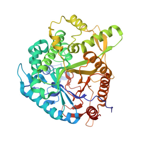
    6YN7

  • PubMed Abstract: 

    β-Glucosidases are used in the food industry to hydrolyse glycosidic bonds in complex sugars, with enzymes sourced from extremophiles better able to tolerate the process conditions. In this work, a novel β-glycosidase from the acidophilic organism Alicyclobacillus herbarius was cloned and heterologously expressed in Escherichia coli BL21(DE3). AheGH1 was stable over a broad range of pH values (5-11) and temperatures (4-55 °C). The enzyme exhibited excellent tolerance to fructose and good tolerance to glucose, retaining 65 % activity in the presence of 10 % (w/v) glucose. It also tolerated organic solvents, some of which appeared to have a stimulating effect, in particular ethanol with a 1.7-fold increase in activity at 10 % (v/v). The enzyme was then applied for the cleavage of isoflavone from isoflavone glucosides in an ethanolic extract of soy flour, to produce soy isoflavones, which constitute a valuable food supplement, full conversion was achieved within 15 min at 30 °C.


  • Organizational Affiliation
    • University of Nottingham, School of Chemistry, Department of Chemical Biology, University Park, Nottingham, NG7 2RD, UK.

Macromolecule Content 

  • Total Structure Weight: 208.54 kDa 
  • Atom Count: 14,649 
  • Modeled Residue Count: 1,774 
  • Deposited Residue Count: 1,808 
  • Unique protein chains: 1

Macromolecules

Find similar proteins by:|  3D Structure
Entity ID: 1
MoleculeChains  Sequence LengthOrganismDetailsImage
AHE, beta-glucosidase enzyme
A, B, C, D
452Paenalicyclobacillus herbariusMutation(s): 0 
EC: 3.2.1.23 (PDB Primary Data), 3.2.1.21 (UniProt)
UniProt
Find proteins for A0A8I3B065 (Alicyclobacillus herbarius)
Explore A0A8I3B065 
Go to UniProtKB:  A0A8I3B065
Entity Groups
Sequence Clusters30% Identity50% Identity70% Identity90% Identity95% Identity100% Identity
UniProt GroupA0A8I3B065
Sequence Annotations
Expand
Reference Sequence

Small Molecules

Ligands 4 Unique
IDChains Name / Formula / InChI Key2D Diagram3D Interactions
PGE
(Subject of Investigation/LOI)

Query on PGE


Download:Ideal Coordinates CCD File
N [auth A],
S [auth B]
TRIETHYLENE GLYCOL
C6 H14 O4
ZIBGPFATKBEMQZ-UHFFFAOYSA-N
PEG
(Subject of Investigation/LOI)

Query on PEG


Download:Ideal Coordinates CCD File
T [auth B]DI(HYDROXYETHYL)ETHER
C4 H10 O3
MTHSVFCYNBDYFN-UHFFFAOYSA-N
EDO
(Subject of Investigation/LOI)

Query on EDO


Download:Ideal Coordinates CCD File
AA [auth D]
BA [auth D]
CA [auth D]
DA [auth D]
E [auth A]
AA [auth D],
BA [auth D],
CA [auth D],
DA [auth D],
E [auth A],
EA [auth D],
F [auth A],
FA [auth D],
G [auth A],
GA [auth D],
H [auth A],
HA [auth D],
I [auth A],
IA [auth D],
J [auth A],
K [auth A],
L [auth A],
O [auth B],
P [auth B],
Q [auth B],
U [auth C],
V [auth C],
W [auth C],
X [auth C],
Y [auth C],
Z [auth D]
1,2-ETHANEDIOL
C2 H6 O2
LYCAIKOWRPUZTN-UHFFFAOYSA-N
NI
(Subject of Investigation/LOI)

Query on NI


Download:Ideal Coordinates CCD File
M [auth A],
R [auth B]
NICKEL (II) ION
Ni
VEQPNABPJHWNSG-UHFFFAOYSA-N

Experimental Data & Validation

Experimental Data

  • Method: X-RAY DIFFRACTION
  • Resolution: 1.98 Å
  • R-Value Free:  0.298 (Depositor), 0.299 (DCC) 
  • R-Value Work:  0.247 (Depositor), 0.247 (DCC) 
  • R-Value Observed: 0.249 (Depositor) 
Space Group: P 1 21 1
Unit Cell:
Length ( Å )Angle ( ˚ )
a = 100.045α = 90
b = 93.35β = 98.692
c = 106.383γ = 90
Software Package:
Software NamePurpose
PHENIXrefinement
XDSdata reduction
STARANISOdata scaling
MOLREPphasing

Structure Validation

View Full Validation Report


Ligand Structure Quality Assessment 


Entry History 

Deposition Data

Revision History  (Full details and data files)

  • Version 1.0: 2021-02-03
    Type: Initial release
  • Version 1.1: 2021-04-14
    Changes: Database references
  • Version 1.2: 2024-01-24
    Changes: Data collection, Database references, Refinement description