6YJY | pdb_00006yjy

Crystal structure of human glutaminyl cyclase in complex with neurotensin 1-5


Experimental Data Snapshot

  • Method: X-RAY DIFFRACTION
  • Resolution: 1.67 Å
  • R-Value Free: 
    0.188 (Depositor), 0.189 (DCC) 
  • R-Value Work: 
    0.169 (Depositor), 0.172 (DCC) 
  • R-Value Observed: 
    0.170 (Depositor) 

Starting Model: experimental
View more details

wwPDB Validation 3D Report Full Report

Validation slider image for 6YJY

Ligand Structure Quality Assessment 


This is version 1.2 of the entry. See complete history

Literature

Hydrazides Are Potent Transition-State Analogues for Glutaminyl Cyclase Implicated in the Pathogenesis of Alzheimer's Disease.

Kupski, O.Funk, L.M.Sautner, V.Seifert, F.Worbs, B.Ramsbeck, D.Meyer, F.Diederichsen, U.Buchholz, M.Schilling, S.Demuth, H.U.Tittmann, K.

(2020) Biochemistry 59: 2585-2591

  • DOI: https://doi.org/10.1021/acs.biochem.0c00337
  • Primary Citation Related Structures: 
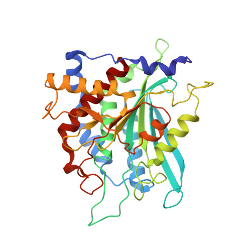
    6YI1, 6YJY

  • PubMed Abstract: 

    Amyloidogenic plaques are hallmarks of Alzheimer's disease (AD) and typically consist of high percentages of modified Aβ peptides bearing N-terminally cyclized glutamate residues. The human zinc(II) enzyme glutaminyl cyclase (QC) was shown in vivo to catalyze the cyclization of N-terminal glutamates of Aβ peptides in a pathophysiological side reaction establishing QC as a druggable target for therapeutic treatment of AD. Here, we report crystallographic snapshots of human QC catalysis acting on the neurohormone neurotensin that delineate the stereochemical course of catalysis and suggest that hydrazides could mimic the transition state of peptide cyclization and deamidation. This hypothesis is validated by a sparse-matrix inhibitor screening campaign that identifies hydrazides as the most potent metal-binding group compared to classic Zn binders. The structural basis of hydrazide inhibition is illuminated by X-ray structure analysis of human QC in complex with a hydrazide-bearing peptide inhibitor and reveals a pentacoordinated Zn complex. Our findings inform novel strategies in the design of potent and highly selective QC inhibitors by employing hydrazides as the metal-binding warhead.


  • Organizational Affiliation
    • Department of Molecular Enzymology, Georg-August University Göttingen, Julia-Lermontowa-Weg 3, 37077 Göttingen, Germany.

Macromolecule Content 

  • Total Structure Weight: 78.41 kDa 
  • Atom Count: 6,161 
  • Modeled Residue Count: 658 
  • Deposited Residue Count: 658 
  • Unique protein chains: 1

Macromolecules

Find similar proteins by:|  3D Structure
Entity ID: 1
MoleculeChains  Sequence LengthOrganismDetailsImage
Glutaminyl-peptide cyclotransferase
A, B
329Homo sapiensMutation(s): 0 
Gene Names: QPCT
EC: 2.3.2.5
UniProt & NIH Common Fund Data Resources
Find proteins for Q16769 (Homo sapiens)
Explore Q16769 
Go to UniProtKB:  Q16769
PHAROS:  Q16769
GTEx:  ENSG00000115828 
Entity Groups
Sequence Clusters30% Identity50% Identity70% Identity90% Identity95% Identity100% Identity
UniProt GroupQ16769
Sequence Annotations
Expand
Reference Sequence

Small Molecules

Ligands 7 Unique
IDChains Name / Formula / InChI Key2D Diagram3D Interactions
MES

Query on MES


Download:Ideal Coordinates CCD File
C [auth A]2-(N-MORPHOLINO)-ETHANESULFONIC ACID
C6 H13 N O4 S
SXGZJKUKBWWHRA-UHFFFAOYSA-N
TYR
(Subject of Investigation/LOI)

Query on TYR


Download:Ideal Coordinates CCD File
BA [auth B],
EA [auth B],
O [auth A]
TYROSINE
C9 H11 N O3
OUYCCCASQSFEME-QMMMGPOBSA-N
GLN
(Subject of Investigation/LOI)

Query on GLN


Download:Ideal Coordinates CCD File
Z [auth B]GLUTAMINE
C5 H10 N2 O3
ZDXPYRJPNDTMRX-VKHMYHEASA-N
LEU
(Subject of Investigation/LOI)

Query on LEU


Download:Ideal Coordinates CCD File
AA [auth B],
DA [auth B],
N [auth A]
LEUCINE
C6 H13 N O2
ROHFNLRQFUQHCH-YFKPBYRVSA-N
PCA
(Subject of Investigation/LOI)

Query on PCA


Download:Ideal Coordinates CCD File
CA [auth B],
M [auth A]
PYROGLUTAMIC ACID
C5 H7 N O3
ODHCTXKNWHHXJC-VKHMYHEASA-N
SO4

Query on SO4


Download:Ideal Coordinates CCD File
D [auth A]
E [auth A]
F [auth A]
G [auth A]
H [auth A]
D [auth A],
E [auth A],
F [auth A],
G [auth A],
H [auth A],
I [auth A],
J [auth A],
K [auth A],
P [auth B],
Q [auth B],
R [auth B],
S [auth B],
T [auth B],
U [auth B],
V [auth B],
W [auth B],
X [auth B]
SULFATE ION
O4 S
QAOWNCQODCNURD-UHFFFAOYSA-L
ZN
(Subject of Investigation/LOI)

Query on ZN


Download:Ideal Coordinates CCD File
L [auth A],
Y [auth B]
ZINC ION
Zn
PTFCDOFLOPIGGS-UHFFFAOYSA-N

Experimental Data & Validation

Experimental Data

  • Method: X-RAY DIFFRACTION
  • Resolution: 1.67 Å
  • R-Value Free:  0.188 (Depositor), 0.189 (DCC) 
  • R-Value Work:  0.169 (Depositor), 0.172 (DCC) 
  • R-Value Observed: 0.170 (Depositor) 
Space Group: H 3 2
Unit Cell:
Length ( Å )Angle ( ˚ )
a = 119.274α = 90
b = 119.274β = 90
c = 334.459γ = 120
Software Package:
Software NamePurpose
XSCALEdata scaling
PHENIXrefinement
PDB_EXTRACTdata extraction
XDSdata reduction
PHASERphasing

Structure Validation

View Full Validation Report


Ligand Structure Quality Assessment 


Entry History 

& Funding Information

Deposition Data


Funding OrganizationLocationGrant Number
German Research Foundation (DFG)GermanyIRTG 1422

Revision History  (Full details and data files)

  • Version 1.0: 2020-07-01
    Type: Initial release
  • Version 1.1: 2020-07-29
    Changes: Database references, Derived calculations
  • Version 1.2: 2024-01-24
    Changes: Data collection, Database references, Refinement description