6YJO | pdb_00006yjo

Structure of FgChi7B


Experimental Data Snapshot

  • Method: X-RAY DIFFRACTION
  • Resolution: 2.38 Å
  • R-Value Free: 
    0.238 (Depositor), 0.238 (DCC) 
  • R-Value Work: 
    0.212 (Depositor), 0.215 (DCC) 
  • R-Value Observed: 
    0.214 (Depositor) 

Starting Model: experimental
View more details

wwPDB Validation 3D Report Full Report

Validation slider image for 6YJO

Ligand Structure Quality Assessment 


This is version 2.4 of the entry. See complete history

Literature

Discovery of fungal oligosaccharide-oxidising flavo-enzymes with previously unknown substrates, redox-activity profiles and interplay with LPMOs.

Haddad Momeni, M.Fredslund, F.Bissaro, B.Raji, O.Vuong, T.V.Meier, S.Nielsen, T.S.Lombard, V.Guigliarelli, B.Biaso, F.Haon, M.Grisel, S.Henrissat, B.Welner, D.H.Master, E.R.Berrin, J.G.Abou Hachem, M.

(2021) Nat Commun 12: 2132-2132

  • DOI: https://doi.org/10.1038/s41467-021-22372-0
  • Primary Citation Related Structures: 
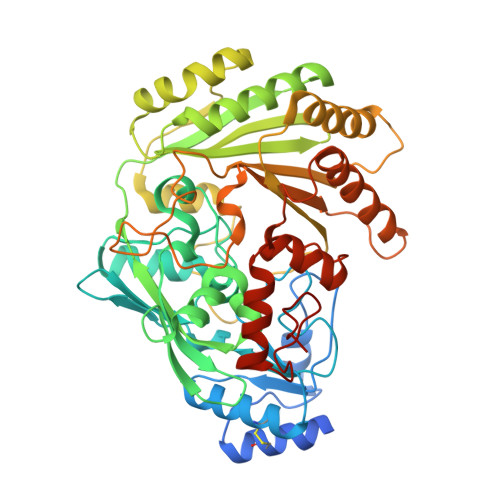
    6YJI, 6YJO

  • PubMed Abstract: 

    Oxidative plant cell-wall processing enzymes are of great importance in biology and biotechnology. Yet, our insight into the functional interplay amongst such oxidative enzymes remains limited. Here, a phylogenetic analysis of the auxiliary activity 7 family (AA7), currently harbouring oligosaccharide flavo-oxidases, reveals a striking abundance of AA7-genes in phytopathogenic fungi and Oomycetes. Expression of five fungal enzymes, including three from unexplored clades, expands the AA7-substrate range and unveils a cellooligosaccharide dehydrogenase activity, previously unknown within AA7. Sequence and structural analyses identify unique signatures distinguishing the strict dehydrogenase clade from canonical AA7 oxidases. The discovered dehydrogenase directly is able to transfer electrons to an AA9 lytic polysaccharide monooxygenase (LPMO) and fuel cellulose degradation by LPMOs without exogenous reductants. The expansion of redox-profiles and substrate range highlights the functional diversity within AA7 and sets the stage for harnessing AA7 dehydrogenases to fine-tune LPMO activity in biotechnological conversion of plant feedstocks.


  • Organizational Affiliation
    • Department of Biotechnology and Biomedicine, Technical University of Denmark, Lyngby, Denmark.

Macromolecule Content 

  • Total Structure Weight: 57.66 kDa 
  • Atom Count: 3,870 
  • Modeled Residue Count: 475 
  • Deposited Residue Count: 512 
  • Unique protein chains: 1

Macromolecules

Find similar proteins by:|  3D Structure
Entity ID: 1
MoleculeChains  Sequence LengthOrganismDetailsImage
FAD-binding PCMH-type domain-containing protein512Fusarium graminearum PH-1Mutation(s): 0 
Gene Names: FGRAMPH1_01T15319
UniProt
Find proteins for A0A098DND1 (Gibberella zeae (strain ATCC MYA-4620 / CBS 123657 / FGSC 9075 / NRRL 31084 / PH-1))
Explore A0A098DND1 
Go to UniProtKB:  A0A098DND1
Entity Groups
Sequence Clusters30% Identity50% Identity70% Identity90% Identity95% Identity100% Identity
UniProt GroupA0A098DND1
Glycosylation
Glycosylation Sites: 1
Sequence Annotations
Expand
Reference Sequence

Oligosaccharides

Help  
Entity ID: 2
MoleculeChains Length2D Diagram GlycosylationD Interactions
beta-D-mannopyranose-(1-4)-2-acetamido-2-deoxy-beta-D-glucopyranose-(1-4)-2-acetamido-2-deoxy-beta-D-glucopyranose
B
3N-Glycosylation
Glycosylation Resources
GlyTouCan: G15407YE
GlyCosmos: G15407YE
GlyGen: G15407YE

Small Molecules

Ligands 1 Unique
IDChains Name / Formula / InChI Key2D Diagram3D Interactions
FAD
(Subject of Investigation/LOI)

Query on FAD


Download:Ideal Coordinates CCD File
C [auth A]FLAVIN-ADENINE DINUCLEOTIDE
C27 H33 N9 O15 P2
VWWQXMAJTJZDQX-UYBVJOGSSA-N

Experimental Data & Validation

Experimental Data

  • Method: X-RAY DIFFRACTION
  • Resolution: 2.38 Å
  • R-Value Free:  0.238 (Depositor), 0.238 (DCC) 
  • R-Value Work:  0.212 (Depositor), 0.215 (DCC) 
  • R-Value Observed: 0.214 (Depositor) 
Space Group: C 1 2 1
Unit Cell:
Length ( Å )Angle ( ˚ )
a = 111.6α = 90
b = 67.455β = 116.31
c = 85.082γ = 90
Software Package:
Software NamePurpose
PHENIXrefinement
XDSdata reduction
XSCALEdata scaling
PHASERphasing

Structure Validation

View Full Validation Report


Ligand Structure Quality Assessment 


Entry History 

& Funding Information

Deposition Data


Funding OrganizationLocationGrant Number
Novo Nordisk FoundationDenmarkNNF17OC0025642

Revision History  (Full details and data files)

  • Version 1.0: 2021-03-03
    Type: Initial release
  • Version 2.0: 2021-03-31
    Changes: Atomic model, Database references, Structure summary
  • Version 2.1: 2021-04-21
    Changes: Database references
  • Version 2.2: 2021-04-28
    Changes: Structure summary
  • Version 2.3: 2024-01-24
    Changes: Data collection, Database references, Refinement description
  • Version 2.4: 2024-10-23
    Changes: Structure summary