


Crystal structure of the first bromodomain of BRD4 in complex with a benzo-diazepine ligand
Picaud, S., Brand, M., Tobias, K., von Delft, F., Arrowsmith, C.H., Edwards, A.M., Bountra, C., Conway, S., Filippakopoulos, P.To be published.
Experimental Data Snapshot
Starting Model: experimental
View more details
Entity ID: 1 | |||||
|---|---|---|---|---|---|
| Molecule | Chains | Sequence Length | Organism | Details | Image |
| Bromodomain-containing protein 4 | 127 | Homo sapiens | Mutation(s): 0 Gene Names: BRD4, HUNK1 | 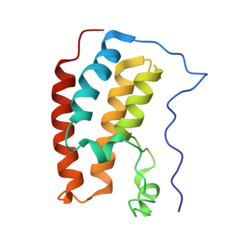 | |
UniProt & NIH Common Fund Data Resources | |||||
PHAROS: O60885 GTEx: ENSG00000141867 | |||||
Entity Groups | |||||
| Sequence Clusters | 30% Identity50% Identity70% Identity90% Identity95% Identity100% Identity | ||||
| UniProt Group | O60885 | ||||
Sequence AnnotationsExpand | |||||
Reference Sequence | |||||
| Ligands 1 Unique | |||||
|---|---|---|---|---|---|
| ID | Chains | Name / Formula / InChI Key | 2D Diagram | 3D Interactions | |
| OS8 (Subject of Investigation/LOI) Download:Ideal Coordinates CCD File | B [auth A] | (4~{R})-~{N}-[3-(7-methoxy-3,4-dihydro-2~{H}-quinolin-1-yl)propyl]-4-methyl-2-oxidanylidene-1,3,4,5-tetrahydro-1,5-benzodiazepine-6-carboxamide C24 H30 N4 O3 XZQSPHYHBYGJPB-MRXNPFEDSA-N |  | ||
| Length ( Å ) | Angle ( ˚ ) |
|---|---|
| a = 41.603 | α = 90 |
| b = 48.347 | β = 90 |
| c = 58.962 | γ = 90 |
| Software Name | Purpose |
|---|---|
| SCALA | data scaling |
| PHASER | phasing |
| REFMAC | refinement |
| PDB_EXTRACT | data extraction |
| XDS | data reduction |
| Funding Organization | Location | Grant Number |
|---|---|---|
| Medical Research Council (MRC, United Kingdom) | United Kingdom | MR/N010051/1 |


