6YGG | pdb_00006ygg

NADase from Aspergillus fumigatus complexed with a substrate anologue


Experimental Data Snapshot

  • Method: X-RAY DIFFRACTION
  • Resolution: 1.85 Å
  • R-Value Free: 
    0.226 (Depositor), 0.203 (DCC) 
  • R-Value Work: 
    0.185 (Depositor) 
  • R-Value Observed: 
    0.187 (Depositor) 

Starting Model: experimental
View more details

wwPDB Validation 3D Report Full Report

Validation slider image for 6YGG

Ligand Structure Quality Assessment 


This is version 1.3 of the entry. See complete history

Literature

Discovery of fungal surface NADases predominantly present in pathogenic species.

Stromland, O.Kallio, J.P.Pschibul, A.Skoge, R.H.Hardardottir, H.M.Sverkeli, L.J.Heinekamp, T.Kniemeyer, O.Migaud, M.Makarov, M.V.Gossmann, T.I.Brakhage, A.A.Ziegler, M.

(2021) Nat Commun 12: 1631-1631

  • DOI: https://doi.org/10.1038/s41467-021-21307-z
  • Primary Citation Related Structures: 
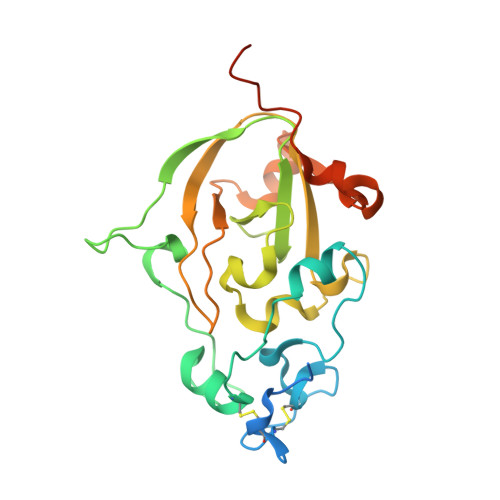
    6YGE, 6YGF, 6YGG

  • PubMed Abstract: 

    Nicotinamide adenine dinucleotide (NAD) is a key molecule in cellular bioenergetics and signalling. Various bacterial pathogens release NADase enzymes into the host cell that deplete the host's NAD + pool, thereby causing rapid cell death. Here, we report the identification of NADases on the surface of fungi such as the pathogen Aspergillus fumigatus and the saprophyte Neurospora crassa. The enzymes harbour a tuberculosis necrotizing toxin (TNT) domain and are predominately present in pathogenic species. The 1.6 Å X-ray structure of the homodimeric A. fumigatus protein reveals unique properties including N-linked glycosylation and a Ca 2+ -binding site whose occupancy regulates activity. The structure in complex with a substrate analogue suggests a catalytic mechanism that is distinct from those of known NADases, ADP-ribosyl cyclases and transferases. We propose that fungal NADases may convey advantages during interaction with the host or competing microorganisms.


  • Organizational Affiliation
    • Department of Biomedicine, University of Bergen, Bergen, Norway.

Macromolecule Content 

  • Total Structure Weight: 59 kDa 
  • Atom Count: 3,998 
  • Modeled Residue Count: 418 
  • Deposited Residue Count: 496 
  • Unique protein chains: 1

Macromolecules

Find similar proteins by:|  3D Structure
Entity ID: 1
MoleculeChains  Sequence LengthOrganismDetailsImage
AfNADase
A, B
248Aspergillus fumigatus Af293Mutation(s): 0 
Gene Names: AFUA_6G14470
EC: 3.2.2 (UniProt), 3.2.2.5 (UniProt)
UniProt
Find proteins for Q4WL81 (Aspergillus fumigatus (strain ATCC MYA-4609 / CBS 101355 / FGSC A1100 / Af293))
Explore Q4WL81 
Go to UniProtKB:  Q4WL81
Entity Groups
Sequence Clusters30% Identity50% Identity70% Identity90% Identity95% Identity100% Identity
UniProt GroupQ4WL81
Glycosylation
Glycosylation Sites: 3
Sequence Annotations
Expand
Reference Sequence

Oligosaccharides

Help  
Entity ID: 2
MoleculeChains Length2D Diagram GlycosylationD Interactions
2-acetamido-2-deoxy-beta-D-glucopyranose-(1-4)-[alpha-L-fucopyranose-(1-6)]2-acetamido-2-deoxy-beta-D-glucopyranose
C, E, F, G
3N-Glycosylation
Glycosylation Resources
GlyTouCan: G21290RB
GlyCosmos: G21290RB
GlyGen: G21290RB
Entity ID: 3
MoleculeChains Length2D Diagram GlycosylationD Interactions
alpha-L-fucopyranose-(1-6)-2-acetamido-2-deoxy-beta-D-glucopyranose
D
2N-Glycosylation
Glycosylation Resources
GlyTouCan: G86851RC
GlyCosmos: G86851RC
GlyGen: G86851RC

Small Molecules

Ligands 4 Unique
IDChains Name / Formula / InChI Key2D Diagram3D Interactions
DQV
(Subject of Investigation/LOI)

Query on DQV


Download:Ideal Coordinates CCD File
K [auth B][(2R,3S,4R,5R)-5-(6-amino-9H-purin-9-yl)-3,4-dihydroxytetrahydrofuran-2-yl]methyl [(2R,3S,4R,5S)-5-(3-carbamoylphenyl)-3,4-dihydroxytetrahydrofuran-2-yl]methyl dihydrogen diphosphate (non-preferred name)
C22 H28 N6 O14 P2
HNKKJJHKTPALEK-RACQCECLSA-N
NAG

Query on NAG


Download:Ideal Coordinates CCD File
H [auth A]2-acetamido-2-deoxy-beta-D-glucopyranose
C8 H15 N O6
OVRNDRQMDRJTHS-FMDGEEDCSA-N
CA
(Subject of Investigation/LOI)

Query on CA


Download:Ideal Coordinates CCD File
I [auth A],
L [auth B]
CALCIUM ION
Ca
BHPQYMZQTOCNFJ-UHFFFAOYSA-N
NA

Query on NA


Download:Ideal Coordinates CCD File
J [auth A]SODIUM ION
Na
FKNQFGJONOIPTF-UHFFFAOYSA-N

Experimental Data & Validation

Experimental Data

  • Method: X-RAY DIFFRACTION
  • Resolution: 1.85 Å
  • R-Value Free:  0.226 (Depositor), 0.203 (DCC) 
  • R-Value Work:  0.185 (Depositor) 
  • R-Value Observed: 0.187 (Depositor) 
Space Group: P 21 21 21
Unit Cell:
Length ( Å )Angle ( ˚ )
a = 51.05α = 90
b = 64.318β = 90
c = 161.007γ = 90
Software Package:
Software NamePurpose
PHENIXrefinement
XDSdata processing
Aimlessdata scaling
PHASERphasing
Cootmodel building
XDSdata reduction

Structure Validation

View Full Validation Report


Ligand Structure Quality Assessment 


Entry History 

Deposition Data

Revision History  (Full details and data files)

  • Version 1.0: 2020-12-23
    Type: Initial release
  • Version 1.1: 2021-07-07
    Changes: Database references
  • Version 1.2: 2024-05-01
    Changes: Data collection, Database references, Refinement description
  • Version 1.3: 2024-10-23
    Changes: Structure summary