6YE8 | pdb_00006ye8

E.coli's Putrescine receptor PotF complexed with Spermidine


Experimental Data Snapshot

  • Method: X-RAY DIFFRACTION
  • Resolution: 1.50 Å
  • R-Value Free: 
    0.183 (Depositor), 0.183 (DCC) 
  • R-Value Work: 
    0.162 (Depositor), 0.163 (DCC) 
  • R-Value Observed: 
    0.162 (Depositor) 

Starting Model: experimental
View more details

wwPDB Validation 3D Report Full Report

Validation slider image for 6YE8

Ligand Structure Quality Assessment 


This is version 1.3 of the entry. See complete history

Literature

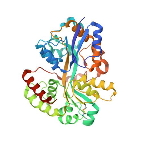
A comprehensive binding study illustrates ligand recognition in the periplasmic binding protein PotF.

Kroger, P.Shanmugaratnam, S.Ferruz, N.Schweimer, K.Hocker, B.

(2021) Structure 29: 433

  • DOI: https://doi.org/10.1016/j.str.2020.12.005
  • Primary Citation Related Structures: 
    6YE0, 6YE6, 6YE7, 6YE8, 6YEB, 6YEC, 6YED

  • PubMed Abstract: 

    Periplasmic binding proteins (PBPs) are ubiquitous receptors in gram-negative bacteria. They sense solutes and play key roles in nutrient uptake. Escherichia coli's putrescine receptor PotF has been reported to bind putrescine and spermidine. We reveal that several similar biogenic polyamines are recognized by PotF. Using isothermal titration calorimetry paired with X-ray crystallography of the different complexes, we unveil PotF's binding modes in detail. The binding site for PBPs is located between two lobes that undergo a large conformational change upon ligand recognition. Hence, analyzing the influence of ligands on complex formation is crucial. Therefore, we solved crystal structures of an open and closed apo state and used them as a basis for molecular dynamics simulations. In addition, we accessed structural behavior in solution for all complexes by 1 H- 15 N HSQC NMR spectroscopy. This combined analysis provides a robust framework for understanding ligand binding for future developments in drug design and protein engineering.


  • Organizational Affiliation
    • Department of Biochemistry, University of Bayreuth, Universitätsstrasse 30, 95447 Bayreuth, Germany.

Macromolecule Content 

  • Total Structure Weight: 80.09 kDa 
  • Atom Count: 6,203 
  • Modeled Residue Count: 682 
  • Deposited Residue Count: 704 
  • Unique protein chains: 1

Macromolecules

Find similar proteins by:|  3D Structure
Entity ID: 1
MoleculeChains  Sequence LengthOrganismDetailsImage
Putrescine-binding periplasmic protein
A, B
352Escherichia coli K-12Mutation(s): 0 
Gene Names: potFb0854JW0838
UniProt
Find proteins for P31133 (Escherichia coli (strain K12))
Explore P31133 
Go to UniProtKB:  P31133
Entity Groups
Sequence Clusters30% Identity50% Identity70% Identity90% Identity95% Identity100% Identity
UniProt GroupP31133
Sequence Annotations
Expand
Reference Sequence

Small Molecules

Ligands 5 Unique
IDChains Name / Formula / InChI Key2D Diagram3D Interactions
SPD
(Subject of Investigation/LOI)

Query on SPD


Download:Ideal Coordinates CCD File
C [auth A],
I [auth B]
SPERMIDINE
C7 H19 N3
ATHGHQPFGPMSJY-UHFFFAOYSA-N
ONT

Query on ONT


Download:Ideal Coordinates CCD File
E [auth A],
L [auth B]
(2~{S})-1-(2-methoxyethoxy)propan-2-amine
C6 H15 N O2
CMXIILNXYHCYPP-LURJTMIESA-N
SO4

Query on SO4


Download:Ideal Coordinates CCD File
O [auth B]SULFATE ION
O4 S
QAOWNCQODCNURD-UHFFFAOYSA-L
JFN

Query on JFN


Download:Ideal Coordinates CCD File
D [auth A]
F [auth A]
G [auth A]
J [auth B]
K [auth B]
D [auth A],
F [auth A],
G [auth A],
J [auth B],
K [auth B],
M [auth B],
N [auth B]
(2R)-1-methoxypropan-2-amine
C4 H11 N O
NXMXETCTWNXSFG-SCSAIBSYSA-N
CL

Query on CL


Download:Ideal Coordinates CCD File
H [auth A],
P [auth B]
CHLORIDE ION
Cl
VEXZGXHMUGYJMC-UHFFFAOYSA-M

Experimental Data & Validation

Experimental Data

  • Method: X-RAY DIFFRACTION
  • Resolution: 1.50 Å
  • R-Value Free:  0.183 (Depositor), 0.183 (DCC) 
  • R-Value Work:  0.162 (Depositor), 0.163 (DCC) 
  • R-Value Observed: 0.162 (Depositor) 
Space Group: P 32 2 1
Unit Cell:
Length ( Å )Angle ( ˚ )
a = 70.83α = 90
b = 70.83β = 90
c = 272.083γ = 120
Software Package:
Software NamePurpose
XDSdata reduction
PHENIXrefinement
XDSdata scaling
Cootmodel building
PHASERphasing

Structure Validation

View Full Validation Report


Ligand Structure Quality Assessment 


Entry History 

& Funding Information

Deposition Data


Funding OrganizationLocationGrant Number
German Research Foundation (DFG)GermanyHO4022/2-3

Revision History  (Full details and data files)

  • Version 1.0: 2021-01-20
    Type: Initial release
  • Version 1.1: 2021-05-19
    Changes: Database references
  • Version 1.2: 2024-01-24
    Changes: Data collection, Database references, Refinement description
  • Version 1.3: 2024-11-06
    Changes: Structure summary