6YAI

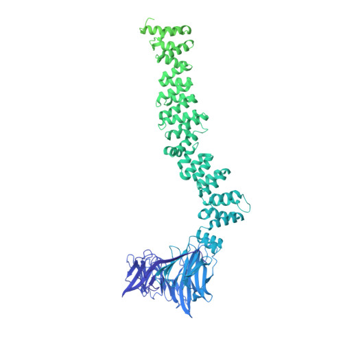
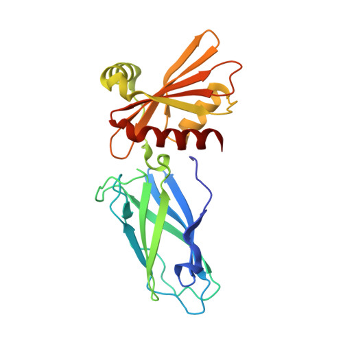
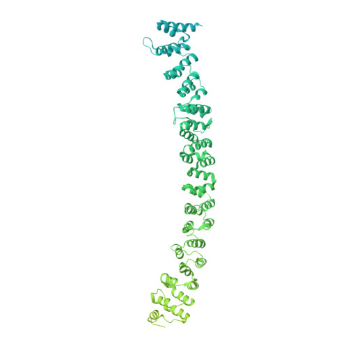
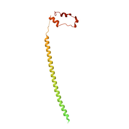
Clathrin with bound beta2 appendage of AP2


Experimental Data Snapshot

  • Method: ELECTRON MICROSCOPY
  • Resolution: 9.20 Å
  • Aggregation State: 3D ARRAY 
  • Reconstruction Method: SUBTOMOGRAM AVERAGING 

Starting Models: experimental
View more details

wwPDB Validation   3D Report Full Report

Currently 6YAI does not have a validation slider image.


This is version 1.3 of the entry. See complete history


Literature

Architecture of the AP2/clathrin coat on the membranes of clathrin-coated vesicles.

Kovtun, O.Dickson, V.K.Kelly, B.T.Owen, D.J.Briggs, J.A.G.

(2020) Sci Adv 6: eaba8381-eaba8381

  • DOI: https://doi.org/10.1126/sciadv.aba8381
  • Primary Citation of Related Structures:  
    6YAE, 6YAF, 6YAH, 6YAI

  • PubMed Abstract: 

    Clathrin-mediated endocytosis (CME) is crucial for modulating the protein composition of a cell's plasma membrane. Clathrin forms a cage-like, polyhedral outer scaffold around a vesicle, to which cargo-selecting clathrin adaptors are attached. Adaptor protein complex (AP2) is the key adaptor in CME. Crystallography has shown AP2 to adopt a range of conformations. Here, we used cryo-electron microscopy, tomography, and subtomogram averaging to determine structures, interactions, and arrangements of clathrin and AP2 at the key steps of coat assembly, from AP2 in solution to membrane-assembled clathrin-coated vesicles (CCVs). AP2 binds cargo and PtdIns(4,5) P 2 (phosphatidylinositol 4,5-bisphosphate)-containing membranes via multiple interfaces, undergoing conformational rearrangement from its cytosolic state. The binding mode of AP2 β2 appendage into the clathrin lattice in CCVs and buds implies how the adaptor structurally modulates coat curvature and coat disassembly.


  • Organizational Affiliation

    MRC Laboratory of Molecular Biology, Cambridge Biomedical Campus, Cambridge CB2 0QH, UK.


Macromolecules
Find similar proteins by:  (by identity cutoff)  |  3D Structure
Entity ID: 1
MoleculeChains Sequence LengthOrganismDetailsImage
Clathrin heavy chain1,630Sus scrofaMutation(s): 0 
UniProt
Find proteins for I3LGD4 (Sus scrofa)
Explore I3LGD4 
Go to UniProtKB:  I3LGD4
Entity Groups  
Sequence Clusters30% Identity50% Identity70% Identity90% Identity95% Identity100% Identity
UniProt GroupI3LGD4
Sequence Annotations
Expand
  • Reference Sequence
Find similar proteins by:  (by identity cutoff)  |  3D Structure
Entity ID: 2
MoleculeChains Sequence LengthOrganismDetailsImage
AP-2 complex subunit betaB [auth F]233Homo sapiensMutation(s): 0 
Gene Names: AP2B1ADTB2CLAPB1
UniProt & NIH Common Fund Data Resources
Find proteins for P63010 (Homo sapiens)
Explore P63010 
Go to UniProtKB:  P63010
PHAROS:  P63010
GTEx:  ENSG00000006125 
Entity Groups  
Sequence Clusters30% Identity50% Identity70% Identity90% Identity95% Identity100% Identity
UniProt GroupP63010
Sequence Annotations
Expand
  • Reference Sequence
Find similar proteins by:  (by identity cutoff)  |  3D Structure
Entity ID: 3
MoleculeChains Sequence LengthOrganismDetailsImage
Clathrin heavy chain1,630Sus scrofaMutation(s): 0 
UniProt
Find proteins for I3LGD4 (Sus scrofa)
Explore I3LGD4 
Go to UniProtKB:  I3LGD4
Entity Groups  
Sequence Clusters30% Identity50% Identity70% Identity90% Identity95% Identity100% Identity
UniProt GroupI3LGD4
Sequence Annotations
Expand
  • Reference Sequence
Find similar proteins by:  (by identity cutoff)  |  3D Structure
Entity ID: 4
MoleculeChains Sequence LengthOrganismDetailsImage
Clathrin light chainG [auth D],
H [auth O],
M [auth I],
N
229Sus scrofaMutation(s): 0 
UniProt
Find proteins for A0A287BFZ5 (Sus scrofa)
Explore A0A287BFZ5 
Go to UniProtKB:  A0A287BFZ5
Entity Groups  
Sequence Clusters30% Identity50% Identity70% Identity90% Identity95% Identity100% Identity
UniProt GroupA0A287BFZ5
Sequence Annotations
Expand
  • Reference Sequence
Experimental Data & Validation

Experimental Data

  • Method: ELECTRON MICROSCOPY
  • Resolution: 9.20 Å
  • Aggregation State: 3D ARRAY 
  • Reconstruction Method: SUBTOMOGRAM AVERAGING 
EM Software:
TaskSoftware PackageVersion
RECONSTRUCTIONTOM
RECONSTRUCTIONAV3
MODEL REFINEMENTPHENIX

Structure Validation

View Full Validation Report

Currently 6YAI does not have a validation slider image.


Entry History & Funding Information

Deposition Data


Funding OrganizationLocationGrant Number
Wellcome TrustUnited Kingdom207455/Z/17/Z
Medical Research Council (MRC, United Kingdom)United KingdomMC_UP_1201/16

Revision History  (Full details and data files)

  • Version 1.0: 2020-07-29
    Type: Initial release
  • Version 1.1: 2020-08-12
    Changes: Database references
  • Version 1.2: 2020-08-19
    Changes: Database references
  • Version 1.3: 2024-05-22
    Changes: Data collection, Database references, Refinement description