


Structure of the human RAB3C in complex with GDP
Diaz-Saez, L., Jung, S., Burgess-Brown, N.A., von Delft, F., Arrowsmith, C.H., Edwards, A., Bountra, C., Huber, K.V.M.To be published.
Experimental Data Snapshot
Starting Model: experimental
View more details
Entity ID: 1 | |||||
|---|---|---|---|---|---|
| Molecule | Chains | Sequence Length | Organism | Details | Image |
| Ras-related protein Rab-3C | 227 | Homo sapiens | Mutation(s): 0 Gene Names: RAB3C EC: 3.6.5.2 | 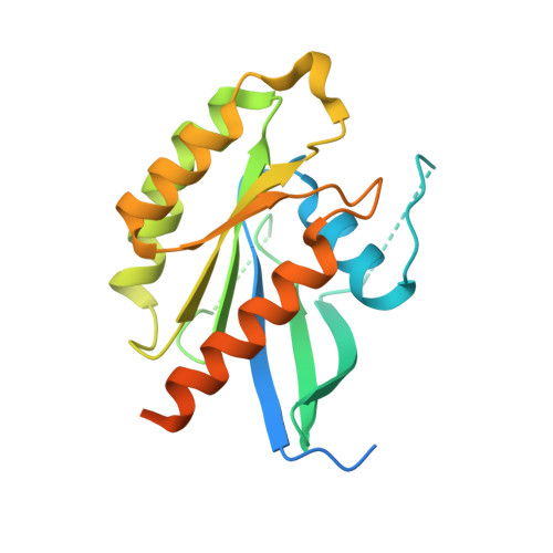 | |
UniProt & NIH Common Fund Data Resources | |||||
PHAROS: Q96E17 GTEx: ENSG00000152932 | |||||
Entity Groups | |||||
| Sequence Clusters | 30% Identity50% Identity70% Identity90% Identity95% Identity100% Identity | ||||
| UniProt Group | Q96E17 | ||||
Sequence AnnotationsExpand | |||||
Reference Sequence | |||||
| Ligands 3 Unique | |||||
|---|---|---|---|---|---|
| ID | Chains | Name / Formula / InChI Key | 2D Diagram | 3D Interactions | |
| GDP (Subject of Investigation/LOI) Download:Ideal Coordinates CCD File | C [auth A], F [auth B] | GUANOSINE-5'-DIPHOSPHATE C10 H15 N5 O11 P2 QGWNDRXFNXRZMB-UUOKFMHZSA-N |  | ||
| BTB Download:Ideal Coordinates CCD File | D [auth A], G [auth B] | 2-[BIS-(2-HYDROXY-ETHYL)-AMINO]-2-HYDROXYMETHYL-PROPANE-1,3-DIOL C8 H19 N O5 OWMVSZAMULFTJU-UHFFFAOYSA-N |  | ||
| MG Download:Ideal Coordinates CCD File | E [auth A], H [auth B] | MAGNESIUM ION Mg JLVVSXFLKOJNIY-UHFFFAOYSA-N |  | ||
| Length ( Å ) | Angle ( ˚ ) |
|---|---|
| a = 40.19 | α = 86.02 |
| b = 40.434 | β = 74.16 |
| c = 66.362 | γ = 66.45 |
| Software Name | Purpose |
|---|---|
| REFMAC | refinement |
| Aimless | data scaling |
| PHASER | phasing |
| PDB_EXTRACT | data extraction |
| DIALS | data reduction |
| Funding Organization | Location | Grant Number |
|---|---|---|
| Innovative Medicines Initiative | -- |


