6XY7 | pdb_00006xy7

Human SHIP1 with magnesium and phosphate bound to the active site


Experimental Data Snapshot

  • Method: X-RAY DIFFRACTION
  • Resolution: 1.09 Å
  • R-Value Free: 
    0.159 (Depositor), 0.159 (DCC) 
  • R-Value Work: 
    0.136 (Depositor), 0.136 (DCC) 

Starting Model: experimental
View more details

wwPDB Validation 3D Report Full Report

Validation slider image for 6XY7

Ligand Structure Quality Assessment 


This is version 1.3 of the entry. See complete history

Literature

Regulation of inositol 5-phosphatase activity by the C2 domain of SHIP1 and SHIP2.

Bradshaw, W.J.Kennedy, E.C.Moreira, T.Smith, L.A.Chalk, R.Katis, V.L.Benesch, J.L.P.Brennan, P.E.Murphy, E.J.Gileadi, O.

(2024) Structure 

  • DOI: https://doi.org/10.1016/j.str.2024.01.005
  • Primary Citation Related Structures: 
    5RW2, 5RW3, 5RW4, 5RW5, 5RW6, 5RW7, 5RW8, 5RW9, 5RWA, 5RWB, 5RWC, 5RWD, 5RWE, 5RWF, 5RWG, 5RWH, 5RWI, 5RWJ, 5RWK, 5RWL, 5RWM, 5RWN, 5RWO, 5RWP, 5RWQ, 5RWR, 5RWS, 5RWT, 5RWU, 5RWV, 5RWW, 5RWX, 5RWY, 5RWZ, 5RX0, 5RX1, 5RX2, 5RX3, 5RX4, 5RX5, 5RX6, 5RX7, 5RX8, 5RX9, 5RXA, 5RXB, 5RXC, 5RXD, 5RXE, 5RXF, ... Search all related entries

  • PubMed Abstract: 

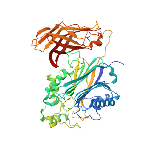
    SHIP1, an inositol 5-phosphatase, plays a central role in cellular signaling. As such, it has been implicated in many conditions. Exploiting SHIP1 as a drug target will require structural knowledge and the design of selective small molecules. We have determined apo, and magnesium and phosphate-bound structures of the phosphatase and C2 domains of SHIP1. The C2 domains of SHIP1 and the related SHIP2 modulate the activity of the phosphatase domain. To understand the mechanism, we performed activity assays, hydrogen-deuterium exchange mass spectrometry, and molecular dynamics on SHIP1 and SHIP2. Our findings demonstrate that the influence of the C2 domain is more pronounced for SHIP2 than SHIP1. We determined 91 structures of SHIP1 with fragments bound, with some near the interface between the two domains. We performed a mass spectrometry screen and determined four structures with covalent fragments. These structures could act as starting points for the development of potent, selective probes.


  • Organizational Affiliation
    • ARUK Oxford Drug Discovery Institute, Centre for Medicines Discovery, Nuffield Department of Medicine Research Building, Old Road Campus, University of Oxford, Oxford OX3 7FZ, UK. Electronic address: william.bradshaw@cmd.ox.ac.uk.

Macromolecule Content 

  • Total Structure Weight: 53.38 kDa 
  • Atom Count: 4,672 
  • Modeled Residue Count: 460 
  • Deposited Residue Count: 463 
  • Unique protein chains: 1

Macromolecules

Find similar proteins by:|  3D Structure
Entity ID: 1
MoleculeChains  Sequence LengthOrganismDetailsImage
Phosphatidylinositol 3,4,5-trisphosphate 5-phosphatase 1A [auth AAA]463Homo sapiensMutation(s): 0 
Gene Names: INPP5DSHIPSHIP1
EC: 3.1.3.86 (PDB Primary Data), 3.1.3.56 (PDB Primary Data), 3.1.3.36 (PDB Primary Data)
UniProt & NIH Common Fund Data Resources
Find proteins for Q92835 (Homo sapiens)
Explore Q92835 
Go to UniProtKB:  Q92835
PHAROS:  Q92835
GTEx:  ENSG00000168918 
Entity Groups
Sequence Clusters30% Identity50% Identity70% Identity90% Identity95% Identity100% Identity
UniProt GroupQ92835
Sequence Annotations
Expand
Reference Sequence

Small Molecules

Ligands 4 Unique
IDChains Name / Formula / InChI Key2D Diagram3D Interactions
PGE

Query on PGE


Download:Ideal Coordinates CCD File
G [auth AAA]TRIETHYLENE GLYCOL
C6 H14 O4
ZIBGPFATKBEMQZ-UHFFFAOYSA-N
PO4
(Subject of Investigation/LOI)

Query on PO4


Download:Ideal Coordinates CCD File
C [auth AAA]PHOSPHATE ION
O4 P
NBIIXXVUZAFLBC-UHFFFAOYSA-K
DMS

Query on DMS


Download:Ideal Coordinates CCD File
D [auth AAA],
E [auth AAA],
F [auth AAA]
DIMETHYL SULFOXIDE
C2 H6 O S
IAZDPXIOMUYVGZ-UHFFFAOYSA-N
MG
(Subject of Investigation/LOI)

Query on MG


Download:Ideal Coordinates CCD File
B [auth AAA]MAGNESIUM ION
Mg
JLVVSXFLKOJNIY-UHFFFAOYSA-N

Experimental Data & Validation

Experimental Data

  • Method: X-RAY DIFFRACTION
  • Resolution: 1.09 Å
  • R-Value Free:  0.159 (Depositor), 0.159 (DCC) 
  • R-Value Work:  0.136 (Depositor), 0.136 (DCC) 
Space Group: P 21 21 21
Unit Cell:
Length ( Å )Angle ( ˚ )
a = 62.5α = 90
b = 79.088β = 90
c = 89.255γ = 90
Software Package:
Software NamePurpose
REFMACrefinement
DIALSdata reduction
STARANISOdata scaling
PHASERphasing

Structure Validation

View Full Validation Report


Ligand Structure Quality Assessment 


Entry History 

& Funding Information

Deposition Data


Funding OrganizationLocationGrant Number
National Institutes of Health/National Institute on Aging (NIH/NIA)United States1U54AG065187-01

Revision History  (Full details and data files)

  • Version 1.0: 2020-02-26
    Type: Initial release
  • Version 1.1: 2020-11-11
    Changes: Source and taxonomy
  • Version 1.2: 2024-01-24
    Changes: Data collection, Database references, Derived calculations, Refinement description
  • Version 1.3: 2024-02-14
    Changes: Database references