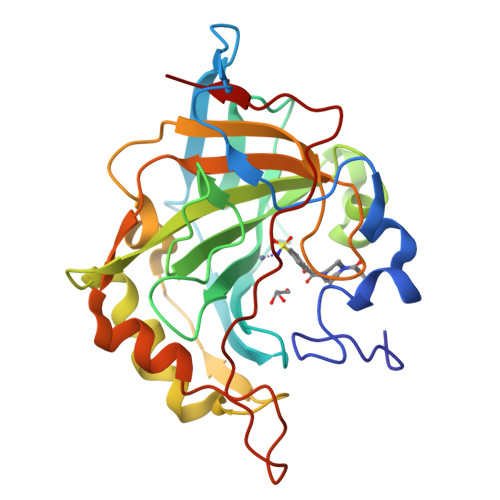
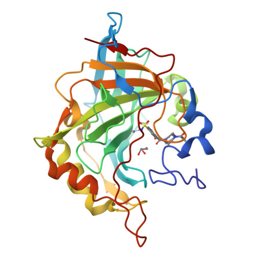
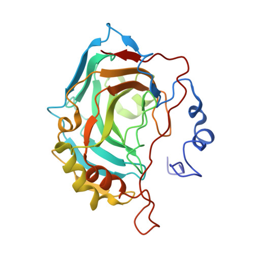
6XXT

The crystal structure of hCA II in complex with a 4-(4-aroylpiperazine-1-carbonyl)benzenesulfonamide derivative.


Experimental Data Snapshot

  • Method: X-RAY DIFFRACTION
  • Resolution: 1.05 Å
  • R-Value Free: 0.145 
  • R-Value Work: 0.135 
  • R-Value Observed: 0.135 

Starting Model: experimental
View more details

wwPDB Validation   3D Report Full Report


Ligand Structure Quality Assessment 

Created with Raphaël 2.3.0Worse 01 BetterLigand structure goodness of fit to experimental dataBest fitted O42Click on this verticalbar to view details

This is version 1.1 of the entry. See complete history


Literature

Looking toward the Rim of the Active Site Cavity of Druggable Human Carbonic Anhydrase Isoforms.

Mancuso, F.Di Fiore, A.De Luca, L.Angeli, A.Monti, S.M.De Simone, G.Supuran, C.T.Gitto, R.

(2020) ACS Med Chem Lett 11: 1000-1005

  • DOI: https://doi.org/10.1021/acsmedchemlett.0c00062
  • Primary Citation of Related Structures:  
    6XXT

  • PubMed Abstract: 

    We report the synthesis and biochemical evaluation of a series of substituted 4-(4-aroylpiperazine-1-carbonyl)benzenesulfonamides ( 5a - s ) developed as inhibitors of druggable carbonic anhydrase (CA) isoforms, as tools for the identification of new therapeutics. X-ray crystallography confirmed that this class of benzenesulfonamides binds CAs through the canonical anchoring of the benzenesulfonamide moiety to the metal ion and a tail-mediated recognition of the middle/top area of the active site cavity. Compound 5e (R = 2-Cl) demonstrated relevant selectivity toward brain-expressed hCA VII. The best balancing in binding affinity and selectivity toward tumor-expressed hCA IX/hCA XII over ubiquitous hCA I/hCA II was found for inhibitor 5o (R = 3-NO 2 ). Notably 5b (R = 2-F) proved to be the most efficacious inhibitor of hCA XII for which computational studies elucidated the CA recognition process.


  • Organizational Affiliation

    Dipartimento di Scienze Chimiche, Biologiche, Farmaceutiche ed Ambientali (CHIBIOFARAM), Università degli Studi di Messina, Viale Palatucci, Polo Didattico SS. Annunziata, 98168 Messina, Italy.


Macromolecules
Find similar proteins by:  (by identity cutoff)  |  3D Structure
Entity ID: 1
MoleculeChains Sequence LengthOrganismDetailsImage
Carbonic anhydrase 2A [auth X]262Homo sapiensMutation(s): 0 
Gene Names: CA2
EC: 4.2.1.1 (PDB Primary Data), 4.2.1.69 (UniProt)
UniProt & NIH Common Fund Data Resources
Find proteins for P00918 (Homo sapiens)
Explore P00918 
Go to UniProtKB:  P00918
PHAROS:  P00918
GTEx:  ENSG00000104267 
Entity Groups  
Sequence Clusters30% Identity50% Identity70% Identity90% Identity95% Identity100% Identity
UniProt GroupP00918
Sequence Annotations
Expand
  • Reference Sequence
Small Molecules
Binding Affinity Annotations 
IDSourceBinding Affinity
O42 BindingDB:  6XXT Ki: 3.7 (nM) from 1 assay(s)
Experimental Data & Validation

Experimental Data

  • Method: X-RAY DIFFRACTION
  • Resolution: 1.05 Å
  • R-Value Free: 0.145 
  • R-Value Work: 0.135 
  • R-Value Observed: 0.135 
  • Space Group: P 1 21 1
Unit Cell:
Length ( Å )Angle ( ˚ )
a = 42.333α = 90
b = 41.455β = 104.26
c = 72.131γ = 90
Software Package:
Software NamePurpose
REFMACrefinement
DENZOdata reduction
SCALEPACKdata scaling
PDB_EXTRACTdata extraction
REFMACphasing

Structure Validation

View Full Validation Report


Ligand Structure Quality Assessment 

Created with Raphaël 2.3.0Worse 01 BetterLigand structure goodness of fit to experimental dataBest fitted O42Click on this verticalbar to view details

Entry History 

Deposition Data

Revision History  (Full details and data files)

  • Version 1.0: 2020-06-03
    Type: Initial release
  • Version 1.1: 2024-01-24
    Changes: Data collection, Database references, Refinement description