6XUI | pdb_00006xui

Crystal structure of human phosphoglucose isomerase in complex with inhibitor


Experimental Data Snapshot

  • Method: X-RAY DIFFRACTION
  • Resolution: 1.95 Å
  • R-Value Free: 
    0.212 (Depositor), 0.217 (DCC) 
  • R-Value Work: 
    0.171 (Depositor), 0.178 (DCC) 
  • R-Value Observed: 
    0.173 (Depositor) 

Starting Model: experimental
View more details

wwPDB Validation 3D Report Full Report

Validation slider image for 6XUI

Ligand Structure Quality Assessment 


This is version 1.1 of the entry. See complete history

Literature

Novel N-substituted 5-phosphate-d-arabinonamide derivatives as strong inhibitors of phosphoglucose isomerases: Synthesis, structure-activity relationship and crystallographic studies.

Ahmad, L.Plancqueel, S.Lazar, N.Korri-Youssoufi, H.Li de la Sierra-Gallay, I.van Tilbeurgh, H.Salmon, L.

(2020) Bioorg Chem 102: 104048-104048

  • DOI: https://doi.org/10.1016/j.bioorg.2020.104048
  • Primary Citation Related Structures: 
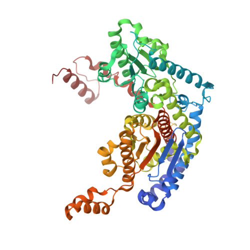
    6XUH, 6XUI

  • PubMed Abstract: 

    Phosphoglucose isomerase (PGI) is a cytosolic enzyme that catalyzes the reversible interconversion of d-glucose 6-phosphate and d-fructose 6-phosphate in glycolysis. Outside the cell, PGI is also known as autocrine motility factor (AMF), a cytokine secreted by a large variety of tumor cells that stimulates motility of cancer cells in vitro and metastases development in vivo. Human PGI and AMF are strictly identical proteins both in terms of sequence and 3D structure, and AMF activity is known to involve, at least in part, the enzymatic active site. Hence, with the purpose of finding new strong AMF-PGI inhibitors that could be potentially used as anticancer agents and/or as bioreceptors for carbohydrate-based electrochemical biosensors, we report in this study the synthesis and kinetic evaluation of several new human PGI inhibitors derived from the synthon 5-phospho-d-arabinono-1,4-lactone. Although not designed as high-energy intermediate analogue inhibitors of the enzyme catalyzed isomerization reaction, several of these N-substituted 5-phosphate-d-arabinonamide derivatives appears as new strong PGI inhibitors. For one of them, we report its crystal structure in complex with human PGI at 2.38 Å. Detailed analysis of its interactions at the active site reveals a new binding mode and shows that human PGI is relatively tolerant for modified inhibitors at the "head" C-1 part, offering promising perspectives for the future design of carbohydrate-based biosensors.


  • Organizational Affiliation
    • Institut de Chimie Moléculaire et des Matériaux d'Orsay (ICMMO), Equipe de Chimie Bioorganique et Bioinorganique, CNRS UMR8182, LabEx LERMIT, Université Paris-Saclay, Rue du Doyen Georges Poitou, bât. 420, 91405 Orsay Cedex, France.

Macromolecule Content 

  • Total Structure Weight: 254.12 kDa 
  • Atom Count: 19,294 
  • Modeled Residue Count: 2,224 
  • Deposited Residue Count: 2,224 
  • Unique protein chains: 1

Macromolecules

Find similar proteins by:|  3D Structure
Entity ID: 1
MoleculeChains  Sequence LengthOrganismDetailsImage
Glucose-6-phosphate isomerase
A, B, C, D
556Homo sapiensMutation(s): 0 
Gene Names: GPI
EC: 5.3.1.9 (PDB Primary Data), 5.1.3 (UniProt)
UniProt & NIH Common Fund Data Resources
Find proteins for P06744 (Homo sapiens)
Explore P06744 
Go to UniProtKB:  P06744
PHAROS:  P06744
GTEx:  ENSG00000105220 
Entity Groups
Sequence Clusters30% Identity50% Identity70% Identity90% Identity95% Identity100% Identity
UniProt GroupP06744
Sequence Annotations
Expand
Reference Sequence

Small Molecules

Ligands 3 Unique
IDChains Name / Formula / InChI Key2D Diagram3D Interactions
PG6

Query on PG6


Download:Ideal Coordinates CCD File
F [auth A],
H [auth B],
J [auth C],
M [auth D]
1-(2-METHOXY-ETHOXY)-2-{2-[2-(2-METHOXY-ETHOXY]-ETHOXY}-ETHANE
C12 H26 O6
DMDPGPKXQDIQQG-UHFFFAOYSA-N
PA5
(Subject of Investigation/LOI)

Query on PA5


Download:Ideal Coordinates CCD File
E [auth A],
G [auth B],
I [auth C],
N [auth D]
5-PHOSPHOARABINONIC ACID
C5 H11 O9 P
HNECGPFIYSOYHF-JJYYJPOSSA-N
GOL

Query on GOL


Download:Ideal Coordinates CCD File
K [auth C],
L [auth C]
GLYCEROL
C3 H8 O3
PEDCQBHIVMGVHV-UHFFFAOYSA-N

Experimental Data & Validation

Experimental Data

  • Method: X-RAY DIFFRACTION
  • Resolution: 1.95 Å
  • R-Value Free:  0.212 (Depositor), 0.217 (DCC) 
  • R-Value Work:  0.171 (Depositor), 0.178 (DCC) 
  • R-Value Observed: 0.173 (Depositor) 
Space Group: P 21 21 21
Unit Cell:
Length ( Å )Angle ( ˚ )
a = 80.551α = 90
b = 106.947β = 90
c = 270.405γ = 90
Software Package:
Software NamePurpose
REFMACrefinement
XDSdata reduction
PDB_EXTRACTdata extraction
XDSdata scaling
PHASERphasing

Structure Validation

View Full Validation Report


Ligand Structure Quality Assessment 


Entry History 

& Funding Information

Deposition Data


Funding OrganizationLocationGrant Number
French National Research AgencyFranceANR-10-LABX-33
French Infrastructure for Integrated Structural Biology (FRISBI)FranceFRISBI ANR-10-INSB-0501

Revision History  (Full details and data files)

  • Version 1.0: 2020-07-29
    Type: Initial release
  • Version 1.1: 2024-01-24
    Changes: Data collection, Database references, Refinement description