6XU3 | pdb_00006xu3

(R)-selective amine transaminase from Shinella sp.


Experimental Data Snapshot

  • Method: X-RAY DIFFRACTION
  • Resolution: 2.10 Å
  • R-Value Free: 
    0.230 (Depositor), 0.230 (DCC) 
  • R-Value Work: 
    0.189 (Depositor), 0.190 (DCC) 
  • R-Value Observed: 
    0.191 (Depositor) 

Starting Model: experimental
View more details

wwPDB Validation 3D Report Full Report

Validation slider image for 6XU3

Ligand Structure Quality Assessment 


This is version 1.2 of the entry. See complete history

Literature

Expanding the Toolbox of R-Selective Amine Transaminases by Identification and Characterization of New Members.

Telzerow, A.Paris, J.Hakansson, M.Gonzalez-Sabin, J.Rios-Lombardia, N.Groger, H.Moris, F.Schurmann, M.Schwab, H.Steiner, K.

(2021) Chembiochem 22: 1232-1242

  • DOI: https://doi.org/10.1002/cbic.202000692
  • Primary Citation Related Structures: 
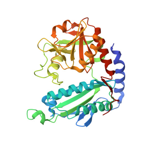
    6SNL, 6XU3

  • PubMed Abstract: 

    Amine transaminases (ATAs) are used to synthesize enantiomerically pure amines, which are building blocks for pharmaceuticals and agrochemicals. R-selective ATAs belong to the fold type IV PLP-dependent enzymes, and different sequence-, structure- and substrate scope-based features have been identified in the past decade. However, our knowledge is still restricted due to the limited number of characterized (R)-ATAs, with additional bias towards fungal origin. We aimed to expand the toolbox of (R)-ATAs and contribute to the understanding of this enzyme subfamily. We identified and characterized four new (R)-ATAs. The ATA from Exophiala sideris contains a motif characteristic for d-ATAs, which was previously believed to be a disqualifying factor for (R)-ATA activity. The crystal structure of the ATA from Shinella is the first from a Gram-negative bacterium. The ATAs from Pseudonocardia acaciae and Tetrasphaera japonica are the first characterized (R)-ATAs with a shortened/missing N-terminal helix. The active-site charges vary significantly between the new and known ATAs, correlating with their diverging substrate scope.


  • Organizational Affiliation
    • Institute of Molecular Biotechnology, Graz University of Technology, Petersgasse 14, 8010, Graz, Austria.

Macromolecule Content 

  • Total Structure Weight: 149.19 kDa 
  • Atom Count: 11,032 
  • Modeled Residue Count: 1,285 
  • Deposited Residue Count: 1,312 
  • Unique protein chains: 1

Macromolecules

Find similar proteins by:|  3D Structure
Entity ID: 1
MoleculeChains  Sequence LengthOrganismDetailsImage
Class IV aminotransferase
A, B, C, D
328Shinella sp. JR1-6Mutation(s): 0 
Gene Names: EXZ48_21850

Small Molecules

Ligands 7 Unique
IDChains Name / Formula / InChI Key2D Diagram3D Interactions
PLG

Query on PLG


Download:Ideal Coordinates CCD File
J [auth B],
N [auth C]
N-GLYCINE-[3-HYDROXY-2-METHYL-5-PHOSPHONOOXYMETHYL-PYRIDIN-4-YL-METHANE]
C10 H15 N2 O7 P
FEVQWBMNLWUBTF-UHFFFAOYSA-N
PLP

Query on PLP


Download:Ideal Coordinates CCD File
E [auth A],
T [auth D]
PYRIDOXAL-5'-PHOSPHATE
C8 H10 N O6 P
NGVDGCNFYWLIFO-UHFFFAOYSA-N
GAB

Query on GAB


Download:Ideal Coordinates CCD File
K [auth B]3-AMINOBENZOIC ACID
C7 H7 N O2
XFDUHJPVQKIXHO-UHFFFAOYSA-N
PEG

Query on PEG


Download:Ideal Coordinates CCD File
I [auth A]DI(HYDROXYETHYL)ETHER
C4 H10 O3
MTHSVFCYNBDYFN-UHFFFAOYSA-N
GOL

Query on GOL


Download:Ideal Coordinates CCD File
G [auth A],
P [auth C],
Q [auth C],
U [auth D],
V [auth D]
GLYCEROL
C3 H8 O3
PEDCQBHIVMGVHV-UHFFFAOYSA-N
NO3

Query on NO3


Download:Ideal Coordinates CCD File
H [auth A]
L [auth B]
M [auth B]
R [auth C]
S [auth C]
H [auth A],
L [auth B],
M [auth B],
R [auth C],
S [auth C],
W [auth D]
NITRATE ION
N O3
NHNBFGGVMKEFGY-UHFFFAOYSA-N
CL

Query on CL


Download:Ideal Coordinates CCD File
F [auth A],
O [auth C]
CHLORIDE ION
Cl
VEXZGXHMUGYJMC-UHFFFAOYSA-M

Experimental Data & Validation

Experimental Data

  • Method: X-RAY DIFFRACTION
  • Resolution: 2.10 Å
  • R-Value Free:  0.230 (Depositor), 0.230 (DCC) 
  • R-Value Work:  0.189 (Depositor), 0.190 (DCC) 
  • R-Value Observed: 0.191 (Depositor) 
Space Group: P 21 21 21
Unit Cell:
Length ( Å )Angle ( ˚ )
a = 75.374α = 90
b = 82.118β = 90
c = 222.435γ = 90
Software Package:
Software NamePurpose
PHENIXrefinement
XDSdata scaling
XDSdata reduction
PHENIXphasing

Structure Validation

View Full Validation Report


Ligand Structure Quality Assessment 


Entry History 

& Funding Information

Deposition Data


Funding OrganizationLocationGrant Number
European CommissionAustria634200

Revision History  (Full details and data files)

  • Version 1.0: 2020-12-09
    Type: Initial release
  • Version 1.1: 2021-04-14
    Changes: Database references
  • Version 1.2: 2024-01-24
    Changes: Data collection, Database references, Refinement description