6XSJ | pdb_00006xsj

X-ray structure of a monoclinic form of alpha amylase from Aspergillus at 1.4 A resolution


Experimental Data Snapshot

  • Method: X-RAY DIFFRACTION
  • Resolution: 1.40 Å
  • R-Value Free: 
    0.237 (Depositor), 0.244 (DCC) 
  • R-Value Work: 
    0.202 (Depositor), 0.209 (DCC) 
  • R-Value Observed: 
    0.204 (Depositor) 

Starting Model: experimental
View more details

wwPDB Validation 3D Report Full Report

Validation slider image for 6XSJ

Ligand Structure Quality Assessment 


This is version 1.4 of the entry. See complete history

Literature

Structures of two novel crystal forms of Aspergillus oryzae alpha amylase (taka-amylase).

Gee, C.L.Holton, J.M.McPherson, A.

(2021) J Biosci Bioeng 131: 605-612

  • DOI: https://doi.org/10.1016/j.jbiosc.2021.02.008
  • Primary Citation Related Structures: 
    6XSJ, 6XSV

  • PubMed Abstract: 

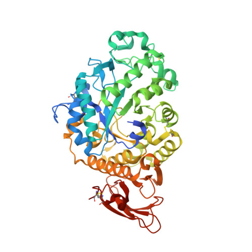
    The structures of Aspergillus oryzae α-amylase were determined in a tetragonal crystal, having one molecule as asymmetric unit, and a monoclinic crystal with two molecules as asymmetric unit. Both crystal forms were obtained from trace contaminants of an old commercial lipase preparation. Structures were determined and refined to 1.65 Å and 1.43 Å resolution respectively. The latter crystal has a non-crystallographic (NCS) twofold axis within the asymmetric unit. Glycosylation at Asn197 is evident, and in the tetragonal crystal can be seen to include three, partially disordered sugar residues following the initial N-acetyl glucosamine (NAG). Superposition of the tetragonal crystal model on the α-amylases from Bacillus subtilis (PDB:1BAG), pig pancreas (PDB:3L2L), and barley (PDB:1AMY), show a high degree of coincidence, particularly for the (β/α) 8 -barrel domains, and especially within the active site. Using this structural agreement between amylases, we extrapolated the binding model of a six residue, limit dextrin found in pig pancreas α-amylase to the A. oryzae enzyme model, which predicts substrate interacting amino acid residues.


  • Organizational Affiliation
    • Department of Molecular and Cell Biology and Howard Hughes Medical Institute, University of California, Stanley Hall 527, Berkeley, CA 94720-3220, USA.

Macromolecule Content 

  • Total Structure Weight: 111.22 kDa 
  • Atom Count: 8,760 
  • Modeled Residue Count: 952 
  • Deposited Residue Count: 998 
  • Unique protein chains: 1

Macromolecules

Find similar proteins by:|  3D Structure
Entity ID: 1
MoleculeChains  Sequence LengthOrganismDetailsImage
Alpha-amylase
A, B
499Aspergillus oryzaeMutation(s): 0 
Gene Names: rhaGamy3OAory_01056410
EC: 3.2.1.1
UniProt
Find proteins for P0C1B3 (Aspergillus oryzae (strain ATCC 42149 / RIB 40))
Explore P0C1B3 
Go to UniProtKB:  P0C1B3
Entity Groups
Sequence Clusters30% Identity50% Identity70% Identity90% Identity95% Identity100% Identity
UniProt GroupP0C1B3
Glycosylation
Glycosylation Sites: 1
Sequence Annotations
Expand
Reference Sequence

Oligosaccharides

Help  
Entity ID: 2
MoleculeChains Length2D Diagram GlycosylationD Interactions
alpha-D-mannopyranose-(1-3)-2-acetamido-2-deoxy-beta-D-glucopyranose
C
2N-Glycosylation
Glycosylation Resources
GlyTouCan: G84445MH
GlyCosmos: G84445MH
GlyGen: G84445MH

Small Molecules

Ligands 7 Unique
IDChains Name / Formula / InChI Key2D Diagram3D Interactions
NAG

Query on NAG


Download:Ideal Coordinates CCD File
E [auth A]2-acetamido-2-deoxy-beta-D-glucopyranose
C8 H15 N O6
OVRNDRQMDRJTHS-FMDGEEDCSA-N
MES

Query on MES


Download:Ideal Coordinates CCD File
G [auth A]2-(N-MORPHOLINO)-ETHANESULFONIC ACID
C6 H13 N O4 S
SXGZJKUKBWWHRA-UHFFFAOYSA-N
PG4

Query on PG4


Download:Ideal Coordinates CCD File
L [auth B]TETRAETHYLENE GLYCOL
C8 H18 O5
UWHCKJMYHZGTIT-UHFFFAOYSA-N
MPD

Query on MPD


Download:Ideal Coordinates CCD File
F [auth A],
I [auth B]
(4S)-2-METHYL-2,4-PENTANEDIOL
C6 H14 O2
SVTBMSDMJJWYQN-YFKPBYRVSA-N
PO4

Query on PO4


Download:Ideal Coordinates CCD File
J [auth B],
K [auth B]
PHOSPHATE ION
O4 P
NBIIXXVUZAFLBC-UHFFFAOYSA-K
ALA

Query on ALA


Download:Ideal Coordinates CCD File
M [auth B]ALANINE
C3 H7 N O2
QNAYBMKLOCPYGJ-REOHCLBHSA-N
CA

Query on CA


Download:Ideal Coordinates CCD File
D [auth A],
H [auth B]
CALCIUM ION
Ca
BHPQYMZQTOCNFJ-UHFFFAOYSA-N

Experimental Data & Validation

Experimental Data

  • Method: X-RAY DIFFRACTION
  • Resolution: 1.40 Å
  • R-Value Free:  0.237 (Depositor), 0.244 (DCC) 
  • R-Value Work:  0.202 (Depositor), 0.209 (DCC) 
  • R-Value Observed: 0.204 (Depositor) 
Space Group: P 1 21 1
Unit Cell:
Length ( Å )Angle ( ˚ )
a = 64.983α = 90
b = 95.189β = 103.51
c = 74.988γ = 90
Software Package:
Software NamePurpose
PHENIXrefinement
PDB_EXTRACTdata extraction
XDSdata reduction
Aimlessdata scaling
PHASERphasing

Structure Validation

View Full Validation Report


Ligand Structure Quality Assessment 


Entry History 

Deposition Data

Revision History  (Full details and data files)

  • Version 1.0: 2020-10-14
    Type: Initial release
  • Version 1.1: 2021-04-21
    Changes: Database references
  • Version 1.2: 2021-06-23
    Changes: Database references
  • Version 1.3: 2023-10-18
    Changes: Advisory, Data collection, Database references, Refinement description
  • Version 1.4: 2024-10-30
    Changes: Structure summary