6XLP

Structure of the essential inner membrane lipopolysaccharide-PbgA complex


Experimental Data Snapshot

  • Method: X-RAY DIFFRACTION
  • Resolution: 2.00 Å
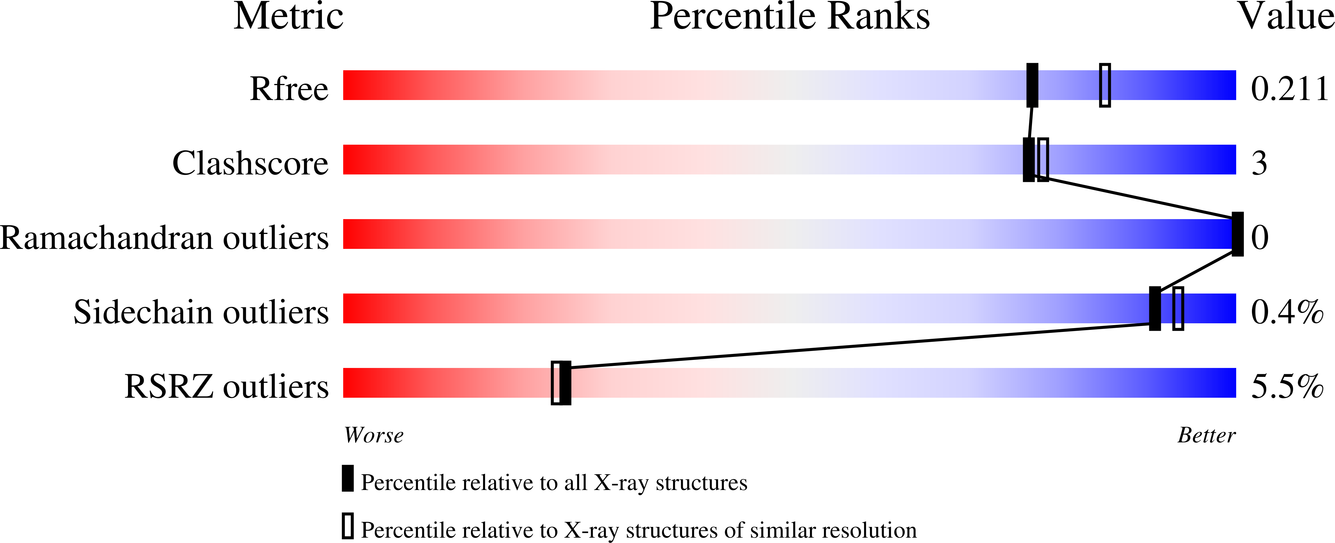
  • R-Value Free: 0.211 
  • R-Value Work: 0.193 
  • R-Value Observed: 0.194 

Starting Model: experimental
View more details

wwPDB Validation   3D Report Full Report


Ligand Structure Quality Assessment 


This is version 1.2 of the entry. See complete history


Literature

Structure of the essential inner membrane lipopolysaccharide-PbgA complex.

Clairfeuille, T.Buchholz, K.R.Li, Q.Verschueren, E.Liu, P.Sangaraju, D.Park, S.Noland, C.L.Storek, K.M.Nickerson, N.N.Martin, L.Dela Vega, T.Miu, A.Reeder, J.Ruiz-Gonzalez, M.Swem, D.Han, G.DePonte, D.P.Hunter, M.S.Gati, C.Shahidi-Latham, S.Xu, M.Skelton, N.Sellers, B.D.Skippington, E.Sandoval, W.Hanan, E.J.Payandeh, J.Rutherford, S.T.

(2020) Nature 584: 479-483

  • DOI: https://doi.org/10.1038/s41586-020-2597-x
  • Primary Citation of Related Structures:  
    6XLP

  • PubMed Abstract: 

    Lipopolysaccharide (LPS) resides in the outer membrane of Gram-negative bacteria where it is responsible for barrier function 1,2 . LPS can cause death as a result of septic shock, and its lipid A core is the target of polymyxin antibiotics 3,4 . Despite the clinical importance of polymyxins and the emergence of multidrug resistant strains 5 , our understanding of the bacterial factors that regulate LPS biogenesis is incomplete. Here we characterize the inner membrane protein PbgA and report that its depletion attenuates the virulence of Escherichia coli by reducing levels of LPS and outer membrane integrity. In contrast to previous claims that PbgA functions as a cardiolipin transporter 6-9 , our structural analyses and physiological studies identify a lipid A-binding motif along the periplasmic leaflet of the inner membrane. Synthetic PbgA-derived peptides selectively bind to LPS in vitro and inhibit the growth of diverse Gram-negative bacteria, including polymyxin-resistant strains. Proteomic, genetic and pharmacological experiments uncover a model in which direct periplasmic sensing of LPS by PbgA coordinates the biosynthesis of lipid A by regulating the stability of LpxC, a key cytoplasmic biosynthetic enzyme 10-12 . In summary, we find that PbgA has an unexpected but essential role in the regulation of LPS biogenesis, presents a new structural basis for the selective recognition of lipids, and provides opportunities for future antibiotic discovery.


  • Organizational Affiliation

    Structural Biology, Genentech Inc., South San Francisco, CA, USA.


Macromolecules
Find similar proteins by:  (by identity cutoff)  |  3D Structure
Entity ID: 1
MoleculeChains Sequence LengthOrganismDetailsImage
LPS binding protein596Escherichia coliMutation(s): 0 
Gene Names: yejMpbgAyejM_2
Membrane Entity: Yes 
UniProt
Find proteins for P0AD27 (Escherichia coli (strain K12))
Explore P0AD27 
Go to UniProtKB:  P0AD27
Entity Groups  
Sequence Clusters30% Identity50% Identity70% Identity90% Identity95% Identity100% Identity
UniProt GroupP0AD27
Sequence Annotations
Expand
  • Reference Sequence
Oligosaccharides

Help

Entity ID: 2
MoleculeChains Length2D Diagram Glycosylation3D Interactions
2-deoxy-3-O-[(1R,3R)-1,3-dihydroxytetradecyl]-2-{[(3R)-3-hydroxytetradecanoyl]amino}-1-O-phosphono-alpha-D-glucopyranose-(6-1)-[3-deoxy-alpha-D-manno-oct-2-ulopyranosonic acid-(2-6)]1,5-anhydro-2-deoxy-2-{[(1S,3R)-1-hydroxy-3-(pentanoyloxy)undecyl]amino}-4-O-phosphono-D-glucitol
B
3N/AN/A
Glycosylation Resources
GlyTouCan:  G54575HS
GlyCosmos:  G54575HS
Small Molecules
Ligands 4 Unique
IDChains Name / Formula / InChI Key2D Diagram3D Interactions
3PE
Query on 3PE

Download Ideal Coordinates CCD File 
V [auth A]1,2-Distearoyl-sn-glycerophosphoethanolamine
C41 H82 N O8 P
LVNGJLRDBYCPGB-LDLOPFEMSA-N
OLB
Query on OLB

Download Ideal Coordinates CCD File 
K [auth A]
L [auth A]
M [auth A]
N [auth A]
O [auth A]
K [auth A],
L [auth A],
M [auth A],
N [auth A],
O [auth A],
P [auth A],
Q [auth A],
R [auth A]
(2S)-2,3-dihydroxypropyl (9Z)-octadec-9-enoate
C21 H40 O4
RZRNAYUHWVFMIP-QJRAZLAKSA-N
PG4
Query on PG4

Download Ideal Coordinates CCD File 
S [auth A],
T [auth A],
U [auth A]
TETRAETHYLENE GLYCOL
C8 H18 O5
UWHCKJMYHZGTIT-UHFFFAOYSA-N
SO4
Query on SO4

Download Ideal Coordinates CCD File 
C [auth A]
D [auth A]
E [auth A]
F [auth A]
G [auth A]
C [auth A],
D [auth A],
E [auth A],
F [auth A],
G [auth A],
H [auth A],
I [auth A],
J [auth A]
SULFATE ION
O4 S
QAOWNCQODCNURD-UHFFFAOYSA-L
Experimental Data & Validation

Experimental Data

  • Method: X-RAY DIFFRACTION
  • Resolution: 2.00 Å
  • R-Value Free: 0.211 
  • R-Value Work: 0.193 
  • R-Value Observed: 0.194 
  • Space Group: C 1 2 1
Unit Cell:
Length ( Å )Angle ( ˚ )
a = 171.272α = 90
b = 56.328β = 106.45
c = 97.359γ = 90
Software Package:
Software NamePurpose
PHENIXrefinement
PDB_EXTRACTdata extraction
HKL-2000data reduction
HKL-2000data scaling
PHASERphasing

Structure Validation

View Full Validation Report


Ligand Structure Quality Assessment 


Entry History 

Deposition Data

Revision History  (Full details and data files)

  • Version 1.0: 2020-08-26
    Type: Initial release
  • Version 1.1: 2020-09-02
    Changes: Database references
  • Version 1.2: 2023-10-18
    Changes: Data collection, Database references, Derived calculations, Refinement description, Structure summary