6XIA | pdb_00006xia

REFINEMENT OF GLUCOSE ISOMERASE FROM STREPTOMYCES ALBUS AT 1.65 ANGSTROMS WITH DATA FROM AN IMAGING PLATE


Experimental Data Snapshot

  • Method: X-RAY DIFFRACTION
  • Resolution: 1.65 Å
  • R-Value Work: 
    0.136 (DCC) 
  • R-Value Observed: 
    0.141 (Depositor) 

wwPDB Validation 3D Report Full Report

Validation slider image for 6XIA

This is version 1.3 of the entry. See complete history

Literature

Refinement of glucose isomerase from Streptomyces albus at 1.65 A with data from an imaging plate.

Dauter, Z.Terry, H.Witzel, H.Wilson, K.S.

(1990) Acta Crystallogr B 46: 833-841

  • DOI: https://doi.org/10.1107/s0108768190008059
  • Primary Citation Related Structures: 
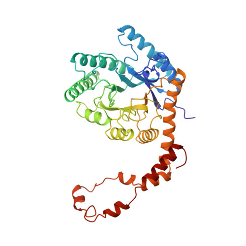
    6XIA

  • PubMed Abstract: 

    The structure of 'metal-free' glucose isomerase of Streptomyces albus strain number YT ATCC 21132 has been analysed and refined at 1.65 A. The space group is I222, with cell dimensions a = 93.9 (1), b = 99.7 (1) and c = 102.9 (1) A, and there is one monomer of the tetrameric molecule per asymmetric unit. The data were recorded from two crystals of the protein using synchrotron radiation from the EMBL beamline X11 at DESY, Hamburg. Data were recorded with an imaging plate scanner designed and built in the EMBL Hamburg outstation. The total data-collection time was less than 12 h and the processing of all data took less than 2 days. The coordinates of the Arthrobacter glucose isomerase refined at a resolution of 2.5 A were used as a starting model. The structure of the protein and of 445 associated water molecules in the asymmetric unit were refined by restrained least-squares minimization using all data between 8 and 1.65 A to a final R factor of 14.1%.


  • Organizational Affiliation
    • European Molecular Biology Laboratory (EMBL), c/o DESY, Hamburg, Federal Republic of Germany.

Macromolecule Content 

  • Total Structure Weight: 43.13 kDa 
  • Atom Count: 3,499 
  • Modeled Residue Count: 387 
  • Deposited Residue Count: 387 
  • Unique protein chains: 1

Macromolecules

Find similar proteins by:|  3D Structure
Entity ID: 1
MoleculeChains  Sequence LengthOrganismDetailsImage
D-XYLOSE ISOMERASE387Streptomyces albusMutation(s): 0 
EC: 5.3.1.5
UniProt
Find proteins for P24299 (Streptomyces albus G)
Explore P24299 
Go to UniProtKB:  P24299
Entity Groups
Sequence Clusters30% Identity50% Identity70% Identity90% Identity95% Identity100% Identity
UniProt GroupP24299
Sequence Annotations
Expand
Reference Sequence

Experimental Data & Validation

Experimental Data

  • Method: X-RAY DIFFRACTION
  • Resolution: 1.65 Å
  • R-Value Work:  0.136 (DCC) 
  • R-Value Observed: 0.141 (Depositor) 
Space Group: I 2 2 2
Unit Cell:
Length ( Å )Angle ( ˚ )
a = 93.9α = 90
b = 99.7β = 90
c = 102.9γ = 90
Software Package:
Software NamePurpose
PROLSQrefinement

Structure Validation

View Full Validation Report


Entry History 

Deposition Data

Revision History  (Full details and data files)

  • Version 1.0: 1991-10-15
    Type: Initial release
  • Version 1.1: 2008-03-25
    Changes: Version format compliance
  • Version 1.2: 2011-07-13
    Changes: Derived calculations, Version format compliance
  • Version 1.3: 2024-03-06
    Changes: Data collection, Database references, Derived calculations, Other