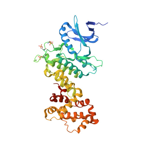
6XDD

Crystal structure of IRE1 in complex with G-3053


Experimental Data Snapshot

  • Method: X-RAY DIFFRACTION
  • Resolution: 2.40 Å
  • R-Value Free: 0.238 
  • R-Value Work: 0.198 
  • R-Value Observed: 0.200 

Starting Model: experimental
View more details

wwPDB Validation   3D Report Full Report


Ligand Structure Quality Assessment 


This is version 1.1 of the entry. See complete history


Literature

Identification of BRaf-Sparing Amino-Thienopyrimidines with Potent IRE1 alpha Inhibitory Activity.

Beveridge, R.E.Wallweber, H.A.Ashkenazi, A.Beresini, M.Clark, K.R.Gibbons, P.Ghiro, E.Kaufman, S.Larivee, A.Leblanc, M.Leclerc, J.P.Lemire, A.Ly, C.Rudolph, J.Schwarz, J.B.Srivastava, S.Wang, W.Zhao, L.Braun, M.G.

(2020) ACS Med Chem Lett 11: 2389-2396

  • DOI: https://doi.org/10.1021/acsmedchemlett.0c00344
  • Primary Citation of Related Structures:  
    6XDB, 6XDD, 6XDF

  • PubMed Abstract: 

    Amino-quinazoline BRaf kinase inhibitor 2 was identified from a library screen as a modest inhibitor of the unfolded protein response (UPR) regulating potential anticancer target IRE1α. A combination of crystallographic and conformational considerations were used to guide structure-based attenuation of BRaf activity and optimization of IRE1α potency. Quinazoline 6-position modifications were found to provide up to 100-fold improvement in IRE1α cellular potency but were ineffective at reducing BRaf activity. A salt bridge contact with Glu651 in IRE1α was then targeted to build in selectivity over BRaf which instead possesses a histidine in this position (His539). Torsional angle analysis revealed that the quinazoline hinge binder core was ill-suited to accommodate the required conformation to effectively reach Glu651, prompting a change to the thienopyrimidine hinge binder. Resulting analogues such as 25 demonstrated good IRE1α cellular potency and imparted more than 1000-fold decrease in BRaf activity.


  • Organizational Affiliation

    Paraza Pharma Inc., 2525 Ave. Marie-Curie, Montreal, QC, Canada H4S 2E1.


Macromolecules
Find similar proteins by:  (by identity cutoff)  |  3D Structure
Entity ID: 1
MoleculeChains Sequence LengthOrganismDetailsImage
Serine/threonine-protein kinase/endoribonuclease IRE1
A, B
434Homo sapiensMutation(s): 0 
Gene Names: ERN1IRE1
EC: 2.7.11.1 (PDB Primary Data), 3.1.26 (PDB Primary Data)
UniProt & NIH Common Fund Data Resources
Find proteins for O75460 (Homo sapiens)
Go to UniProtKB:  O75460
PHAROS:  O75460
GTEx:  ENSG00000178607 
Sequence Annotations
Expand
  • Reference Sequence
Small Molecules
Ligands 1 Unique
IDChains Name / Formula / InChI Key2D Diagram3D Interactions
N97 (Subject of Investigation/LOI)
Query on N97

Download Ideal Coordinates CCD File 
C [auth A],
D [auth B]
4-[(trans-4-aminocyclohexyl)amino]-N-(6-chloro-3-{[(2,5-difluorophenyl)sulfonyl]amino}-2-fluorophenyl)thieno[3,2-d]pyrimidine-7-carboxamide
C25 H22 Cl F3 N6 O3 S2
NALUYXQOXMHJMM-HDJSIYSDSA-N
Modified Residues  1 Unique
IDChains TypeFormula2D DiagramParent
SEP
Query on SEP
A, B
L-PEPTIDE LINKINGC3 H8 N O6 PSER
Binding Affinity Annotations 
IDSourceBinding Affinity
N97 BindingDB:  6XDD IC50: min: 24, max: 320 (nM) from 2 assay(s)
Experimental Data & Validation

Experimental Data

  • Method: X-RAY DIFFRACTION
  • Resolution: 2.40 Å
  • R-Value Free: 0.238 
  • R-Value Work: 0.198 
  • R-Value Observed: 0.200 
  • Space Group: P 21 21 21
Unit Cell:
Length ( Å )Angle ( ˚ )
a = 47.201α = 90
b = 156.816β = 90
c = 157.766γ = 90
Software Package:
Software NamePurpose
Aimlessdata scaling
PHENIXrefinement
PDB_EXTRACTdata extraction
XDSdata reduction
PHASERphasing

Structure Validation

View Full Validation Report


Ligand Structure Quality Assessment 


Entry History 

Deposition Data

Revision History  (Full details and data files)

  • Version 1.0: 2021-04-21
    Type: Initial release
  • Version 1.1: 2023-10-18
    Changes: Data collection, Database references, Refinement description