6XCJ | pdb_00006xcj

Crystal Structure of DH650 Fab from a Rhesus Macaque in Complex with HIV-1 gp120 Core


Experimental Data Snapshot

  • Method: X-RAY DIFFRACTION
  • Resolution: 2.80 Å
  • R-Value Free: 
    0.284 (Depositor), 0.297 (DCC) 
  • R-Value Work: 
    0.227 (Depositor), 0.233 (DCC) 
  • R-Value Observed: 
    0.230 (Depositor) 

Starting Model: experimental
View more details

wwPDB Validation 3D Report Full Report

Validation slider image for 6XCJ

Ligand Structure Quality Assessment 


This is version 1.3 of the entry. See complete history

Literature

Recapitulation of HIV-1 Env-antibody coevolution in macaques leading to neutralization breadth.

Roark, R.S.Li, H.Williams, W.B.Chug, H.Mason, R.D.Gorman, J.Wang, S.Lee, F.H.Rando, J.Bonsignori, M.Hwang, K.K.Saunders, K.O.Wiehe, K.Moody, M.A.Hraber, P.T.Wagh, K.Giorgi, E.E.Russell, R.M.Bibollet-Ruche, F.Liu, W.Connell, J.Smith, A.G.DeVoto, J.Murphy, A.I.Smith, J.Ding, W.Zhao, C.Chohan, N.Okumura, M.Rosario, C.Ding, Y.Lindemuth, E.Bauer, A.M.Bar, K.J.Ambrozak, D.Chao, C.W.Chuang, G.Y.Geng, H.Lin, B.C.Louder, M.K.Nguyen, R.Zhang, B.Lewis, M.G.Raymond, D.D.Doria-Rose, N.A.Schramm, C.A.Douek, D.C.Roederer, M.Kepler, T.B.Kelsoe, G.Mascola, J.R.Kwong, P.D.Korber, B.T.Harrison, S.C.Haynes, B.F.Hahn, B.H.Shaw, G.M.

(2021) Science 371

  • DOI: https://doi.org/10.1126/science.abd2638
  • Primary Citation Related Structures: 
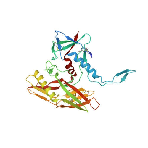
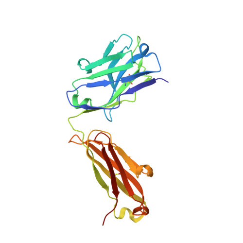
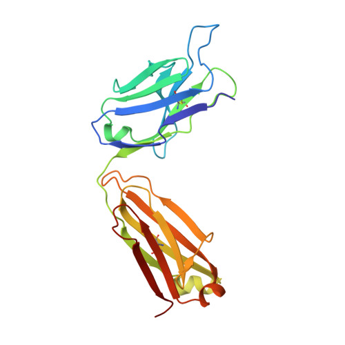
    6XCJ, 6XRT

  • PubMed Abstract: 

    Neutralizing antibodies elicited by HIV-1 coevolve with viral envelope proteins (Env) in distinctive patterns, in some cases acquiring substantial breadth. We report that primary HIV-1 envelope proteins-when expressed by simian-human immunodeficiency viruses in rhesus macaques-elicited patterns of Env-antibody coevolution very similar to those in humans, including conserved immunogenetic, structural, and chemical solutions to epitope recognition and precise Env-amino acid substitutions, insertions, and deletions leading to virus persistence. The structure of one rhesus antibody, capable of neutralizing 49% of a 208-strain panel, revealed a V2 apex mode of recognition like that of human broadly neutralizing antibodies (bNAbs) PGT145 and PCT64-35S. Another rhesus antibody bound the CD4 binding site by CD4 mimicry, mirroring human bNAbs 8ANC131, CH235, and VRC01. Virus-antibody coevolution in macaques can thus recapitulate developmental features of human bNAbs, thereby guiding HIV-1 immunogen design.


  • Organizational Affiliation
    • Departments of Medicine and Microbiology, Perelman School of Medicine, University of Pennsylvania, Philadelphia, PA 19104, USA.

Macromolecule Content 

  • Total Structure Weight: 90.13 kDa 
  • Atom Count: 5,712 
  • Modeled Residue Count: 721 
  • Deposited Residue Count: 803 
  • Unique protein chains: 3

Macromolecules

Find similar proteins by:|  3D Structure
Entity ID: 1
MoleculeChains  Sequence LengthOrganismDetailsImage
Envelope Glycoprotein gp120A [auth G]357Human immunodeficiency virus 1Mutation(s): 0 
UniProt
Find proteins for A0A0A7I3C6 (Human immunodeficiency virus type 1)
Explore A0A0A7I3C6 
Go to UniProtKB:  A0A0A7I3C6
Entity Groups
Sequence Clusters30% Identity50% Identity70% Identity90% Identity95% Identity100% Identity
UniProt GroupA0A0A7I3C6
Glycosylation
Glycosylation Sites: 9
Sequence Annotations
Expand
Reference Sequence
Find similar proteins by:|  3D Structure
Entity ID: 2
MoleculeChains  Sequence LengthOrganismDetailsImage
DH650 Fab Heavy ChainB [auth H]226Macaca mulattaMutation(s): 0 
Find similar proteins by:|  3D Structure
Entity ID: 3
MoleculeChains  Sequence LengthOrganismDetailsImage
DH650 Fab Light ChainC [auth L]220Macaca mulattaMutation(s): 0 

Small Molecules

Ligands 1 Unique
IDChains Name / Formula / InChI Key2D Diagram3D Interactions
NAG

Query on NAG


Download:Ideal Coordinates CCD File
D [auth G]
E [auth G]
F [auth G]
G
H [auth G]
D [auth G],
E [auth G],
F [auth G],
G,
H [auth G],
I [auth G],
J [auth G],
K [auth G],
L [auth G]
2-acetamido-2-deoxy-beta-D-glucopyranose
C8 H15 N O6
OVRNDRQMDRJTHS-FMDGEEDCSA-N

Experimental Data & Validation

Experimental Data

  • Method: X-RAY DIFFRACTION
  • Resolution: 2.80 Å
  • R-Value Free:  0.284 (Depositor), 0.297 (DCC) 
  • R-Value Work:  0.227 (Depositor), 0.233 (DCC) 
  • R-Value Observed: 0.230 (Depositor) 
Space Group: P 21 21 2
Unit Cell:
Length ( Å )Angle ( ˚ )
a = 138.52α = 90
b = 122.1β = 90
c = 53.4γ = 90
Software Package:
Software NamePurpose
BUSTERrefinement
XDSdata reduction
XSCALEdata scaling
PHASERphasing

Structure Validation

View Full Validation Report


Ligand Structure Quality Assessment 


Entry History 

& Funding Information

Deposition Data


Funding OrganizationLocationGrant Number
National Institutes of Health/National Institute Of Allergy and Infectious Diseases (NIH/NIAID)United StatesAI144371

Revision History  (Full details and data files)

  • Version 1.0: 2020-12-02
    Type: Initial release
  • Version 1.1: 2021-01-20
    Changes: Database references
  • Version 1.2: 2023-10-18
    Changes: Data collection, Database references, Refinement description
  • Version 1.3: 2024-11-06
    Changes: Structure summary