6XCG

Histone-lysine N-methyltransferase NSD2-PWWP1 with compound UNC6934


Experimental Data Snapshot

  • Method: X-RAY DIFFRACTION
  • Resolution: 1.64 Å
  • R-Value Free: 0.222 
  • R-Value Work: 0.178 
  • R-Value Observed: 0.179 

Starting Model: experimental
View more details

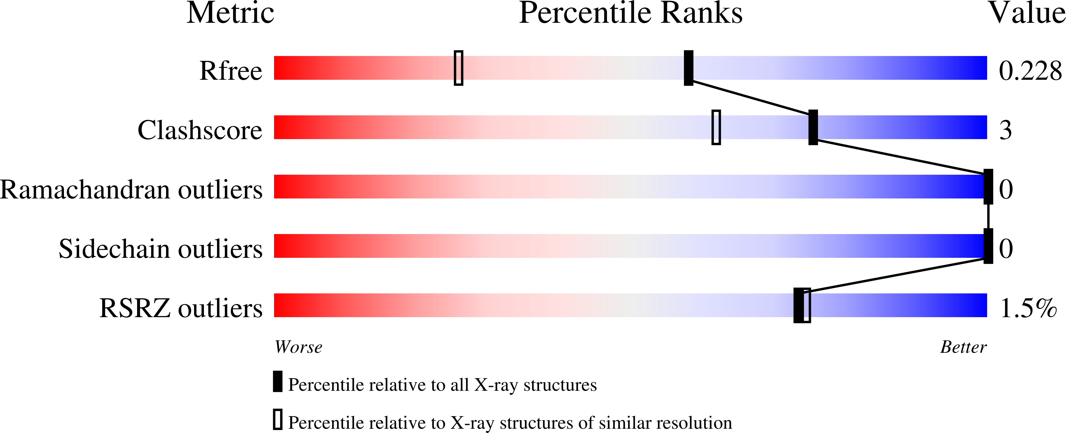
wwPDB Validation   3D Report Full Report


Ligand Structure Quality Assessment 

Created with Raphaël 2.3.0Worse 01 BetterLigand structure goodness of fit to experimental dataBest fitted V01Click on this verticalbar to view details

This is version 1.2 of the entry. See complete history


Literature

A chemical probe targeting the PWWP domain alters NSD2 nucleolar localization.

Dilworth, D.Hanley, R.P.Ferreira de Freitas, R.Allali-Hassani, A.Zhou, M.Mehta, N.Marunde, M.R.Ackloo, S.Carvalho Machado, R.A.Khalili Yazdi, A.Owens, D.D.G.Vu, V.Nie, D.Y.Alqazzaz, M.Marcon, E.Li, F.Chau, I.Bolotokova, A.Qin, S.Lei, M.Liu, Y.Szewczyk, M.M.Dong, A.Kazemzadeh, S.Abramyan, T.Popova, I.K.Hall, N.W.Meiners, M.J.Cheek, M.A.Gibson, E.Kireev, D.Greenblatt, J.F.Keogh, M.C.Min, J.Brown, P.J.Vedadi, M.Arrowsmith, C.H.Barsyte-Lovejoy, D.James, L.I.Schapira, M.

(2022) Nat Chem Biol 18: 56-63

  • DOI: https://doi.org/10.1038/s41589-021-00898-0
  • Primary Citation of Related Structures:  
    6XCG, 7MDN

  • PubMed Abstract: 

    Nuclear receptor-binding SET domain-containing 2 (NSD2) is the primary enzyme responsible for the dimethylation of lysine 36 of histone 3 (H3K36), a mark associated with active gene transcription and intergenic DNA methylation. In addition to a methyltransferase domain, NSD2 harbors two proline-tryptophan-tryptophan-proline (PWWP) domains and five plant homeodomains (PHDs) believed to serve as chromatin reading modules. Here, we report a chemical probe targeting the N-terminal PWWP (PWWP1) domain of NSD2. UNC6934 occupies the canonical H3K36me2-binding pocket of PWWP1, antagonizes PWWP1 interaction with nucleosomal H3K36me2 and selectively engages endogenous NSD2 in cells. UNC6934 induces accumulation of endogenous NSD2 in the nucleolus, phenocopying the localization defects of NSD2 protein isoforms lacking PWWP1 that result from translocations prevalent in multiple myeloma (MM). Mutations of other NSD2 chromatin reader domains also increase NSD2 nucleolar localization and enhance the effect of UNC6934. This chemical probe and the accompanying negative control UNC7145 will be useful tools in defining NSD2 biology.


  • Organizational Affiliation

    Structural Genomics Consortium, University of Toronto, Toronto, Ontario, Canada. david.dilworth@utoronto.ca.


Macromolecules
Find similar proteins by:  (by identity cutoff)  |  3D Structure
Entity ID: 1
MoleculeChains Sequence LengthOrganismDetailsImage
Histone-lysine N-methyltransferase NSD2
A, B, C
141Homo sapiensMutation(s): 0 
Gene Names: NSD2KIAA1090MMSETTRX5WHSC1
EC: 2.1.1.356 (PDB Primary Data), 2.1.1.357 (UniProt)
UniProt & NIH Common Fund Data Resources
Find proteins for O96028 (Homo sapiens)
Explore O96028 
Go to UniProtKB:  O96028
PHAROS:  O96028
GTEx:  ENSG00000109685 
Entity Groups  
Sequence Clusters30% Identity50% Identity70% Identity90% Identity95% Identity100% Identity
UniProt GroupO96028
Sequence Annotations
Expand
  • Reference Sequence
Small Molecules
Ligands 2 Unique
IDChains Name / Formula / InChI Key2D Diagram3D Interactions
V01 (Subject of Investigation/LOI)
Query on V01

Download Ideal Coordinates CCD File 
D [auth A],
E [auth B],
I [auth C]
N-cyclopropyl-3-oxo-N-({4-[(pyrimidin-4-yl)carbamoyl]phenyl}methyl)-3,4-dihydro-2H-1,4-benzoxazine-7-carboxamide
C24 H21 N5 O4
KOZGEDUWAQFVAV-UHFFFAOYSA-N
UNX
Query on UNX

Download Ideal Coordinates CCD File 
F [auth B]
G [auth B]
H [auth B]
J [auth C]
K [auth C]
UNKNOWN ATOM OR ION
X
Binding Affinity Annotations 
IDSourceBinding Affinity
V01 BindingDB:  6XCG Kd: min: 91, max: 500 (nM) from 2 assay(s)
Experimental Data & Validation

Experimental Data

  • Method: X-RAY DIFFRACTION
  • Resolution: 1.64 Å
  • R-Value Free: 0.222 
  • R-Value Work: 0.178 
  • R-Value Observed: 0.179 
  • Space Group: P 1
Unit Cell:
Length ( Å )Angle ( ˚ )
a = 49.566α = 91.35
b = 49.978β = 91.88
c = 51.179γ = 118.27
Software Package:
Software NamePurpose
REFMACrefinement
SCALEPACKdata scaling
PHASERphasing
PDB_EXTRACTdata extraction
DENZOdata reduction

Structure Validation

View Full Validation Report


Ligand Structure Quality Assessment 

Created with Raphaël 2.3.0Worse 01 BetterLigand structure goodness of fit to experimental dataBest fitted V01Click on this verticalbar to view details

Entry History 

Revision History  (Full details and data files)

  • Version 1.0: 2020-07-22
    Type: Initial release
  • Version 1.1: 2023-08-23
    Changes: Data collection, Database references, Refinement description
  • Version 1.2: 2023-10-18
    Changes: Refinement description