6XB4 | pdb_00006xb4

Structure of PrGV poxin in post-reactive state with Gp[2'-5']Ap[3']


Experimental Data Snapshot

  • Method: X-RAY DIFFRACTION
  • Resolution: 1.90 Å
  • R-Value Free: 
    0.207 (Depositor), 0.214 (DCC) 
  • R-Value Work: 
    0.183 (Depositor) 
  • R-Value Observed: 
    0.183 (Depositor) 

wwPDB Validation 3D Report Full Report

Validation slider image for 6XB4

Ligand Structure Quality Assessment 


This is version 1.1 of the entry. See complete history

Literature

Structures of diverse poxin cGAMP nucleases reveal a widespread role for cGAS-STING evasion in host-pathogen conflict.

Eaglesham, J.B.McCarty, K.L.Kranzusch, P.J.

(2020) Elife 9

  • DOI: https://doi.org/10.7554/eLife.59753
  • Primary Citation Related Structures: 
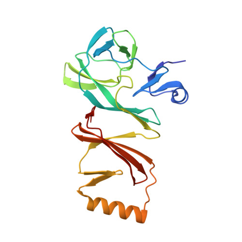
    6XB3, 6XB4, 6XB5, 6XB6

  • PubMed Abstract: 

    DNA viruses in the family Poxviridae encode poxin enzymes that degrade the immune second messenger 2'3'-cGAMP to inhibit cGAS-STING immunity in mammalian cells. The closest homologs of poxin exist in the genomes of insect viruses suggesting a key mechanism of cGAS-STING evasion may have evolved outside of mammalian biology. Here we use a biochemical and structural approach to discover a broad family of 369 poxins encoded in diverse viral and animal genomes and define a prominent role for 2'3'-cGAMP cleavage in metazoan host-pathogen conflict. Structures of insect poxins reveal unexpected homology to flavivirus proteases and enable identification of functional self-cleaving poxins in RNA-virus polyproteins. Our data suggest widespread 2'3'-cGAMP signaling in insect antiviral immunity and explain how a family of cGAS-STING evasion enzymes evolved from viral proteases through gain of secondary nuclease activity. Poxin acquisition by poxviruses demonstrates the importance of environmental connections in shaping evolution of mammalian pathogens.


  • Organizational Affiliation
    • Department of Microbiology, Harvard Medical School, Boston, United States.

Macromolecule Content 

  • Total Structure Weight: 94.21 kDa 
  • Atom Count: 7,103 
  • Modeled Residue Count: 765 
  • Deposited Residue Count: 800 
  • Unique protein chains: 1

Macromolecules

Find similar proteins by:|  3D Structure
Entity ID: 1
MoleculeChains  Sequence LengthOrganismDetailsImage
Poxin
A, B, C, D
200Pieris rapae granulovirusMutation(s): 0 
UniProt
Find proteins for A0ACD6B8Q5 (Pieris rapae granulovirus)
Explore A0ACD6B8Q5 
Go to UniProtKB:  A0ACD6B8Q5
Entity Groups
Sequence Clusters30% Identity50% Identity70% Identity90% Identity95% Identity100% Identity
UniProt GroupA0ACD6B8Q5
Sequence Annotations
Expand
Reference Sequence

Experimental Data & Validation

Experimental Data

  • Method: X-RAY DIFFRACTION
  • Resolution: 1.90 Å
  • R-Value Free:  0.207 (Depositor), 0.214 (DCC) 
  • R-Value Work:  0.183 (Depositor) 
  • R-Value Observed: 0.183 (Depositor) 
Space Group: P 1 21 1
Unit Cell:
Length ( Å )Angle ( ˚ )
a = 77.47α = 90
b = 58.14β = 108.548
c = 100.698γ = 90
Software Package:
Software NamePurpose
PHENIXrefinement
XDSdata reduction
Aimlessdata scaling
AutoSolphasing

Structure Validation

View Full Validation Report


Ligand Structure Quality Assessment 


Entry History 

Deposition Data

Revision History  (Full details and data files)

  • Version 1.0: 2020-11-25
    Type: Initial release
  • Version 1.1: 2024-03-06
    Changes: Data collection, Database references