6XAI | pdb_00006xai

Crystal structure of NzeB in complex with cyclo-(L-Trp-L-Pro)


Experimental Data Snapshot

  • Method: X-RAY DIFFRACTION
  • Resolution: 1.49 Å
  • R-Value Free: 
    0.212 (Depositor), 0.213 (DCC) 
  • R-Value Work: 
    0.167 (Depositor), 0.171 (DCC) 
  • R-Value Observed: 
    0.169 (Depositor) 

Starting Model: in silico
View more details

wwPDB Validation 3D Report Full Report

Validation slider image for 6XAI

Ligand Structure Quality Assessment 


This is version 1.3 of the entry. See complete history

Literature

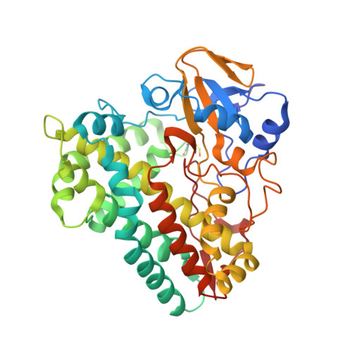
Structure and Function of NzeB, a Versatile C-C and C-N Bond-Forming Diketopiperazine Dimerase.

Shende, V.V.Khatri, Y.Newmister, S.A.Sanders, J.N.Lindovska, P.Yu, F.Doyon, T.J.Kim, J.Houk, K.N.Movassaghi, M.Sherman, D.H.

(2020) J Am Chem Soc 142: 17413-17424

  • DOI: https://doi.org/10.1021/jacs.0c06312
  • Primary Citation Related Structures: 
    6XAI, 6XAJ, 6XAK, 6XAL, 6XAM

  • PubMed Abstract: 

    The dimeric diketopiperazine (DKPs) alkaloids are a diverse family of natural products (NPs) whose unique structural architectures and biological activities have inspired the development of new synthetic methodologies to access these molecules. However, catalyst-controlled methods that enable the selective formation of constitutional and stereoisomeric dimers from a single monomer are lacking. To resolve this long-standing synthetic challenge, we sought to characterize the biosynthetic enzymes that assemble these NPs for application in biocatalytic syntheses. Genome mining enabled identification of the cytochrome P450, NzeB ( Streptomyces sp. NRRL F-5053), which catalyzes both intermolecular carbon-carbon (C-C) and carbon-nitrogen (C-N) bond formation. To identify the molecular basis for the flexible site-selectivity, stereoselectivity, and chemoselectivity of NzeB, we obtained high-resolution crystal structures (1.5 Å) of the protein in complex with native and non-native substrates. This, to our knowledge, represents the first crystal structure of an oxidase catalyzing direct, intermolecular C-H amination. Site-directed mutagenesis was utilized to assess the role individual active-site residues play in guiding selective DKP dimerization. Finally, computational approaches were employed to evaluate plausible mechanisms regarding NzeB function and its ability to catalyze both C-C and C-N bond formation. These results provide a structural and computational rationale for the catalytic versatility of NzeB, as well as new insights into variables that control selectivity of CYP450 diketopiperazine dimerases.


  • Organizational Affiliation
    • Department of Chemistry and Biochemistry, University of California, Los Angeles, California 90095, United States.

Macromolecule Content 

  • Total Structure Weight: 90.29 kDa 
  • Atom Count: 6,989 
  • Modeled Residue Count: 798 
  • Deposited Residue Count: 802 
  • Unique protein chains: 1

Macromolecules

Find similar proteins by:|  3D Structure
Entity ID: 1
MoleculeChains  Sequence LengthOrganismDetailsImage
NzeB
A, B
401Streptomyces sp. NRRL F-5053Mutation(s): 0 
UniProt
Find proteins for A0A8I3B043 (Streptomyces sp. NRRL F-5053)
Explore A0A8I3B043 
Go to UniProtKB:  A0A8I3B043
Entity Groups
Sequence Clusters30% Identity50% Identity70% Identity90% Identity95% Identity100% Identity
UniProt GroupA0A8I3B043
Sequence Annotations
Expand
Reference Sequence

Small Molecules

Ligands 3 Unique
IDChains Name / Formula / InChI Key2D Diagram3D Interactions
HEM
(Subject of Investigation/LOI)

Query on HEM


Download:Ideal Coordinates CCD File
C [auth A],
I [auth B]
PROTOPORPHYRIN IX CONTAINING FE
C34 H32 Fe N4 O4
KABFMIBPWCXCRK-RGGAHWMASA-L
QRP
(Subject of Investigation/LOI)

Query on QRP


Download:Ideal Coordinates CCD File
D [auth A],
E [auth A],
J [auth B],
K [auth B]
(3S,8aS)-3-(1H-indol-3-ylmethyl)hexahydropyrrolo[1,2-a]pyrazine-1,4-dione
C16 H17 N3 O2
RYFZBPVMVYTEKZ-KBPBESRZSA-N
EDO

Query on EDO


Download:Ideal Coordinates CCD File
F [auth A],
G [auth A],
H [auth A],
L [auth B],
M [auth B]
1,2-ETHANEDIOL
C2 H6 O2
LYCAIKOWRPUZTN-UHFFFAOYSA-N
Modified Residues  1 Unique
IDChains TypeFormula2D DiagramParent
CSO
Query on CSO
A, B
L-PEPTIDE LINKINGC3 H7 N O3 SCYS

Experimental Data & Validation

Experimental Data

  • Method: X-RAY DIFFRACTION
  • Resolution: 1.49 Å
  • R-Value Free:  0.212 (Depositor), 0.213 (DCC) 
  • R-Value Work:  0.167 (Depositor), 0.171 (DCC) 
  • R-Value Observed: 0.169 (Depositor) 
Space Group: P 1
Unit Cell:
Length ( Å )Angle ( ˚ )
a = 56.122α = 91.13
b = 56.285β = 92.93
c = 58.779γ = 101
Software Package:
Software NamePurpose
PHENIXrefinement
PDB_EXTRACTdata extraction
XDSdata reduction
XDSdata scaling
PHENIXphasing

Structure Validation

View Full Validation Report


Ligand Structure Quality Assessment 


Entry History 

& Funding Information

Deposition Data


Funding OrganizationLocationGrant Number
National Science Foundation (NSF, United States)United StatesCHE-1700982
National Institutes of Health/National Institute of General Medical Sciences (NIH/NIGMS)United StatesR35-GM118101

Revision History  (Full details and data files)

  • Version 1.0: 2021-06-09
    Type: Initial release
  • Version 1.1: 2021-12-22
    Changes: Database references
  • Version 1.2: 2024-04-03
    Changes: Data collection, Refinement description
  • Version 1.3: 2024-10-23
    Changes: Structure summary